Showing posts with label Dentistry Open Access Journals. Show all posts

Thursday, February 27, 2020

Lupine Publishers | Bioactivity, Biocompatibility and Biomimetic Properties for Dental Materials: Clarifying the Confusion?

Lupine Publishers | Journal of Oral Healthcare

Abstract

Often in the profession of dentistry, a new or novel instrument, material, technique, and/or “system” is introduced which can incur a “state-of-the-art” status without necessarily being subjected to the rigors of clinical testing or longitudinal patient-based studies prior to receiving the stamp of approval or the moniker of “standard of care”. Recently, provocative terminology surrounding the field of dental materials has been publicized through the literature, promoting exciting claims and possible long-term advancements for patient care. In this “new era” of evidence-based restorative dentistry; conservative interdiction, i.e. “informed” removal of diseased tissue with concurrent substitution considering form and function, esthetics, and the interaction of the physical and mechanical properties of the replacement materials with living, dynamic structures found in the human tooth, has been of paramount importance.
Abbrevations: ACP: Amorphous Calcium Phosphates, MTA: Mineral Trioxide Aggregate, PVPA: Poly Vinyl Phosponic Acid, PAA: Polyacrylic Acids

Introduction

The progression or evolution of dentistry has occurred, to a great degree, in concert, with the development of material technology [1]. During the last two decades, the categorization of dental materials, specifically, adhesive systems and composite resins have included the term “nanotechnology” into the lexicon of scientific literature [2]. Nanotechnology involves the science and engineering of functional molecules at the nanoscale (onebillionth of a meter) level [2]. As applied to dentistry, this innovative approach promotes the incorporation or interaction of nanostructured materials together with the complex arrangement of organic/inorganic molecular-level constituents comprising living tooth structure, allowing for a myriad of possible preventive and therapeutic applications [2]. Owing to this progression of material development, the assignments of additional revolutionary dimensions have included the origination of the concepts of biocompatibility or bioactivity into dental science.
As a possible expansion of nanotechnology applied to dental materials: the terms biocompatible, bioactive, bioinduction, and biomimetics can be defined independently; however, have often been characterized synonymously [3]. Biocompatible is simply a term to describe a substance or material that will do no harm to existing living structures, that is non-mutagenic and noncytotoxic. The term “bioactivity” was first described in 1969 by Hench, whereby a “bioactive material is one that elicits a specific biological response at the interface of the material which results in the formation of a bond between the tissues and the material” [4]. Furthermore, the definition was refined and updated to include two categories based upon intent and procedure, originally pertaining, specifically, to bone tissue:
a) Class A: A material that elicits an intracellular and extracellular response (osteoproductive);
b) Class B: Materials eliciting an extracellular response only (osteocontuctive) [5].

Accordingly, a bioactive material can have “the effect on, or eliciting a response from living tissue, organisms, or cells”, thus contributing to the formation of a new substance or creation of a living, compatible system [3]. A bioinductive property is defined as “the capability of a material for inducing a response in a biologic system”[3]. Biomimetics is the “study of formation, structure, or function of biologically produced substances and materials and biological mechanisms and processes for the purpose of synthesizing similar products by artificial mechanisms that mimic natural substances”[3,6]. So, although these terms seem to imply different connotations, what can a dental practitioner conclude, deduce, and/or apply for everyday use? Any substance, arrived from by any process (bioactive, bioinduction, biomimetic) should exhibit attributes of being biocompatible. It appears that both a bioactive and biomimetic substance can include the process of bioinduction and that a biomimetic substance could possibly be produced through bioactive activities.

Bioactive materials and processes are probably the most applicable for endodontics and restorative dentistry based upon current uses: luting cements, pulp capping agents, root repair materials, permanent restorations, hard tissue remineralization (fluoride, calcium, and phosphate ions) and bone regeneration properties, and treatment of dentinal hypersensitivity[1,3,7-13]. In order for these materials to become biocompatibily active or retain characteristics of bioactivity; bactericidal and bacteriostatic (inhibits bacterial growth and biofilm formation) properties for the stimulation of reparative dentin formation and maintenance of pulpal vitality must be achieved and maintained [3]. Examples include fluorides for remineralization, antibacterial resins and cements (Reactimer bond™ Shofu Dental Corp., Kyoto, Japan; ABF™ Kuraray, Kurasiki, Japan), restoratives (Active™ BioACTIVE, PULPDENT Corp., Watertown, MA, USA) releasing fluorides and containing amorphous calcium phosphates [ACP], medicaments (mineral trioxide aggregate [MTA] and bioaggregate; Biodentine™, Septodont, Lancaster, PA, USA; TheraCal™, Bisco Dental Products, Schaumburg, ILL, USA; and Endosequence root repair [RRM]™, Brasseler USA, Savannah, GA), and luting cements (Ceramir Crown & Bridge, Doxa Dental Inc., Chicago, ILL, USA) that induce healing and/or for creation of new tooth structures[1,3,7,8,10-14]. Biomimetic substances include the usage of polyvinylphosponic acid (PVPA) polyacrylic acids (PAA) as calcium phosphate matrix protein analogues for remineralization purposes [7,15].

Conclusion

Although these materials are in their infancy, with long-term efficacy based on improvements of mechanical and physical properties pending, future materials will hopefully create circumstances for increased tooth-like attributes due to properties of adhesion, remineralization, and integration [1,3,7].

Read more Lupine Publishers Blogger Articles please click on: https://lupinepublishers.blogspot.com/


Follow on Twitter       : https://twitter.com/lupine_online 

Monday, February 24, 2020

Lupine Publishers | Down’s Syndrome- A Disease Caused By Genetic Alteration

Lupine Publishers | Dentistry Open Access Journal

Abstract

Down’s syndrome is the most common syndrome, medical professional encounters in day to day practice. It is a genetic disorder with a typical face profile and few classical intraoral features. Herein we report case and review on Down’s syndrome with facial features.
Keywords: Down’s Syndrome; Trisomy; Chromosome; Oral Manifestation

Introduction

Down syndrome is one of the commonest disorders with huge medical and social cost. DS is associated with number of phenotypes including congenital heart defects, leukemia, Alzheimer’s disease, Hirsch sprung disease etc. [1]. Down syndrome is a prevalent genetic disorder in intellectual disability in India. Its prevalence in tribal population is not known [2]. Down syndrome is one of the leading genetic causes of intellectual disability in the world. DS alone accounts 15-20% of ID population across the world [3,4].

Case Report

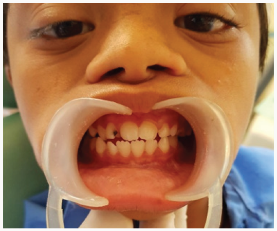
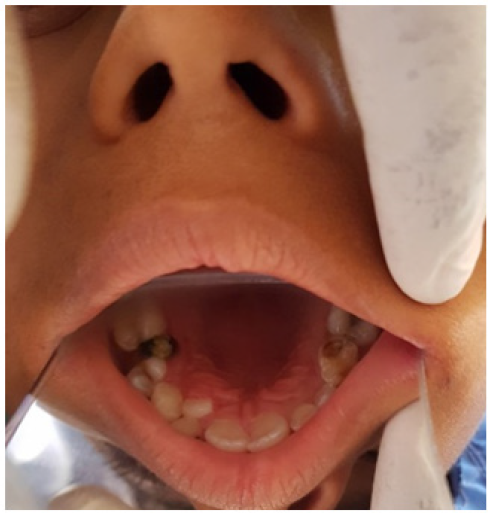
An 8 year old male patient came to the department of oral medicine and radiology for routine dental check-up. Extra oral examination revealed characteristic facial profile with increased inter canthal distance (Figure 1). Intraoral examination revealed Gingiva was soft with deposits on the teeth, High arched palate, with depressed nasal bridge was seen (Figure 2). Macro glossia was also seen .Correlating the intraoral and extra oral findings a Provisional diagnosis of Down’s syndrome/ Trisomy 21 was given. Patient was referred to the respective departments of pedodontics for restoration of decayed teeth.]
Figure 1: Extra oral features showing increased inter canthal distance and depressed nasal bridge.
Lupinepublishers-openaccess-dentistry-oral-healthcare
Figure 2: Intraoral features showing high arched palate.
Lupinepublishers-openaccess-dentistry-oral-healthcare

Discussion

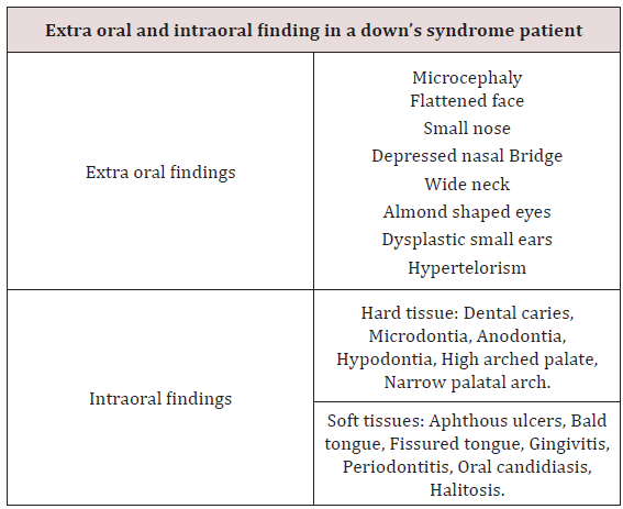
Down syndrome is one of the most leading causes of intellectual disability and millions of these patients face various health issues including learning and memory, congenital heart diseases, Alzheimer’s diseases, leukemia, cancers and Hirsch rung disease. The incidence of trisomy is influenced by maternal age and differs in population [5,6]. Facial findings in the patients can be characterised into extra oral and intraoral features (Table 1) [7]. Parents of children with Down’s syndrome should be aware of these possible conditions so they can be diagnosed and treated quickly and appropriately. According to Asim A et al. A Down’s syndrome child should have regular check-up from various consultants. These include:
a) Clinical geneticist - Referral to a genetic counselling program is highly desirable.
b) Developmental paediatrician.
c) Cardiologist - Early cardiologic evaluation is crucial for diagnosing and treating congenital heart defects, which occur in as many as 60% of these patients.
d) Paediatric pneumonologist -Recurrent respiratory tract infections are common in patients with DS.
e) Ophthalmologist.
f) Neurologist/Neurosurgeon - As many as 10% of patients with DS have epilepsy; therefore, neurologic evaluation may be needed.
g) Orthopaedic specialist.
h) Child psychiatrist - A child psychiatrist should lead liaison interventions, family therapies, and psychometric evaluations.
i) Physical and occupational therapist.
j) Speech-language pathologist.
k) Audiologist.
l) Paediatric dentist.
Hackshaw AK et al in their study, proposed a new screening method in which measurements obtained during 1st and 2nd trimester are integrated to provide the risk status of having pregnancy with DS. Moderate to severe intellectual disability occur as a constant feature, with IQ’s ranging from 20 to 85 [8]. Kennard in his review stated that there are a number of ultrasound markers in Down’s syndrome which includes nuchal fold thickness, cardiac abnormalities, duodenal atresia, femur length & pyelectasis [9]. The signs and symptoms of Down’s syndrome are characterised by neotenization of brain and bodies. Management strategies such as early childhood intervention, screening from common problems, medical treatment when indicated, a conductive family environment and vocational training can improve the overall development of children with Down’s syndrome [10].
Table 1:
Lupinepublishers-openaccess-dentistry-oral-healthcare

Conclusion

Genetics have always have played a major role in physical and mental being of an individual. Downs patients being mentally and medically weak, best care needs to be taken with adequate precautions.

Read more Lupine Publishers Blogger Articles please click on: https://lupinepublishers.blogspot.com/


Follow on Twitter       : https://twitter.com/lupine_online

Thursday, February 6, 2020

Lupine Publishers | Caregiver’s Oral Healthcare Practices And The Level of Utilisation Of Oral Health Services and The Dental Caries Experience Of 3-12-Year-Olds Suffering From Heart Disease in Nairobi, Kenya

Lupine Publishers | Dental and Oral Health Journals

Abstract

Cardiac diseases require that there is the meticulous maintenance of oral hygiene to avoid bacteremia, which has been associated with rheumatic heart disease and bacterial endocarditis. The aim was to establish the utilisation of oral health care and oral health practices of the caregiver about the oral hygiene and caries experience of children aged 3-12 years suffering from heart disease and were attending three pediatric cardiology clinics in Nairobi, Kenya. The study was descriptive and cross-sectional. It involved a study sample of children suffering from different types of cardiac conditions and attending the Pediatric cardiac clinics in three public institutions in Nairobi Kenya. The instruments the caregivers used to brush the children’s teeth were the toothbrush 61(75%); chewing stick 14(17%) and 6 (8%) never cleaned their teeth. Children who used a chewing stick had a lower dmft of 1.40±2.98 compared to a dmft of 3.22±3.59 among children who used the toothbrush, with Mann Whitney U, Z p=0.024 (p≤0.05).The children who brushed their teeth had a lower mean plaque score of 1.68±0.58 compared those who did not clean with a mean plaque of 2.28±0.40 with a Mann Whitney U, Z=-2.611, p=0.009(p≤0.05). It was noted that the children who had visited a dentist had a higher caries experience with a dmft of 4.18±4.13 and DMFT of 1.16±1.92. However, the children who had never sought treatment at a dental facility had lower dmft of 1.89±2.88; and DMFT of 0.36±1, and the differences were statistically significant with Mann Whitney U, Z p=0.008(p≤0.05). The plaque scores and caries experience were high in children whose caregivers had low aggregate utilisation of the oral health care facilities. However, those who had a low aggregate of oral hygiene practices had slightly higher plaque scores and caries experience.
Keywords: Cardiac Disease; Children; Utilisation; Oral Health Services; Caregivers

Introduction

Populations with chronic medical illness or other disabilities had the most unmet needs for oral health services [1], with poor oral hygiene and increased caries experience than the general population. For a child from a low-income family with heart disease, this means an added economic burden in an already tricky situation [2], as heart diseases necessitate regular dental check-ups and maintenance of meticulous oral hygiene. This concern has even been highlighted with new proposals on changes in the guidelines relating to prophylaxis against infective endocarditis [3,4]. The oral conditions may have a considerable impact on the general health status and quality of life of otherwise healthy children, but their effects on those children with acute and chronic illness can be more dangerous [5]. Children with cardiac defects and diseases are at increased risk or even life-threatening complications [6]. Hence the need for preventive dental health care geared to reducing the risks associated with management of the oral conditions under general anaesthesia. Also, the prolonged bleeding from warfarin medication often taken By the children [7-10]. Poor oral hygiene may give rise to a frequent bacteraemia under normal physiological conditions, and this can lead to a permanent risk of developing heart disease [11-14]. Two common oral diseases, namely periodontal and dental caries, though preventable, are still more prevalent in Kenya [15,16]. The children with heart disease have the disadvantage that their caregivers are preoccupied with the with the primary medical condition the cardiac disease, resulting in the neglect of other facets of the child’s total health [17]. The Kenya National Oral Health policy document has already indicated that the dmft value for Kenyan 5-year old children as at 2002 was 1.5±2.2, while 43% of 6-8-year-old children had caries [15], underscoring the fact that caries is still very rampant amongst the child population in Kenya.
The study was descriptive and cross-sectional where all the patients aged 3 to 12 years and their caregivers attending paediatric cardiology clinics over a three month period at Kenyatta National Hospital (KNH), Gertrude’s Garden Children’s Hospital (GGCH) and Mater Hospital. A Purposive sampling had been used to select the study hospitals. Based on Kliegman. study, the study population sample was determined as 79 cases. However, 81 patients were recruited in the study. A semi-structured questionnaire was used to collect information on the socio-demographic characteristics of the children and the parent/guardian habits on oral health practices and utilization of oral health services. As children waited to consult the cardiologist clinical examinations done to record the oral health status. The examination was conducted using sterilized instruments and under natural daylight, with the participants seated on a chair facing the window. Great care was taken during periodontal probing for gingivitis, to avoid initiating bleeding that could lead to septicaemia as the children were not on prophylactic antibiotics. The results were recorded on predesigned individual questionnaire sheets, and a record of dental caries and plaque was done. The dental caries was then recorded as dmft for the primary dentition and DMFT in the permanent [18,19], and the dental plaque was marked based on the Loe and Silness plaque score index [20]. Before commencement of the study, the examiner was calibrated by an experienced paediatric dentist on the collection of data relating to dental caries, and dental plaque Cohen’s kappa index score of 0.87 and 0.85 (n=10) was obtained for dental caries and plaque score respectively. The questionnaire was pre-tested before use. A duplicate clinical examination was also performed by the examiner to determine intra-examiner consistency, with results of Cohen’s kappa index score of 0.91 and 0.86 (n=12) being obtained for dental caries and plaque score respectively.

Data analysis

The data collected was cleaned, coded and analyzed using SPSS version 17-computer software from SPSS Inc. IL. The results obtained were compared and tested using Kruskal Wallis Chi-square and Mann Whitney U statistical tests, with statistical significance pegged at 95% confidence interval.

Results

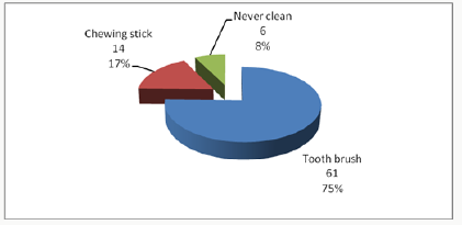
The 81 children in the study, 44 (54.3%) were males and 37 (45.7%) females. Their ages ranged between 3-12 years with a mean age of 8.16 years (± 2.81 SD), and the 6-9-year-olds accounted for the most substantial proportion of 33 (40.7%) compared to the 3-5 year-olds who formed 16(19.8%). The differences in ages and gender were not statistically significant Chi χ2 =1.287, two df, p=0.525(p≤0.05). A total of 37(46%) children were from rural areas, 28(34%) were from Nairobi, and 16(20%) were from other urban centres other than Nairobi. The distribution of the children according to the type of heart disease, rheumatic (RHD) accounted for 36(44.5%) while infective endocarditis (IE) affected 4(4.9%). The duration since diagnosis of the cardiopathy ranged from less than one year to 12 years. Nearly half of the children, 40 (49%) had been diagnosed with the disease for a duration of between 1 to 5 years, while those who had been diagnosed more than five years and those less than one year accounted for 30% and 21% respectively. The caregivers’ oral health care practices that included how the child’s teeth were brushed; the frequency of brushing; and whether tooth brushing was supervised showed that 75(93%) children cleaned their teeth and 6(7%) children did not clean their teeth. Of the group that cleaned their teeth, 33(44%) did it twice a day, 29(39%) once a day while 16% once in a while/occasionally. About supervision, 62 (83%) reported cleaning their teeth without supervision while 13 were assisted by the caregivers. Inquiry on the ways the child’s teeth were cleaned, 75% (61) of the children used toothbrush and the rest of the results were as shown in Figure 1. The children who used toothpaste were 59 (79%) while 16 (21%) never use any toothpaste.
Figure 1:
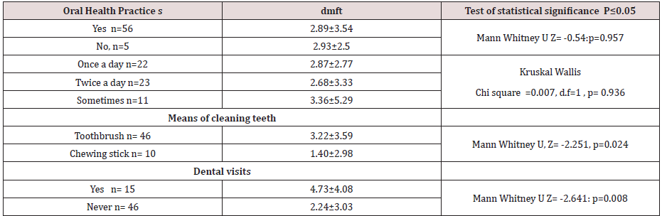
Considering the utilisation of oral health care services by children with heart diseases; fifty-nine (72.8%), children had never visited a dentist or utilised oral health services. Among the 22 (27.2%) children who had been to a dentist, the dental procedure during the last appointment included extraction 10 (12.3%). Also cleaning/prophylaxis (1(1.2%)), consultation ; check-up 9(11.1%) and fillings 2(2.5%).Caregiver’s oral healthcare practices and the dental caries experience about the children five children who never cleaned their teeth had a higher dmft of 2.93±2.50 compared to a lower dmft of 2.89 ±3.54 among the 56 children who cleaned their teeth, and the differences were insignificant with p=0.957(p≤0.05).
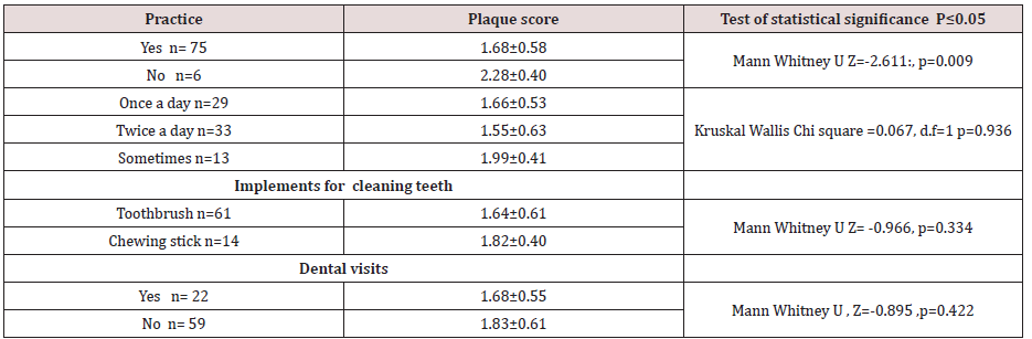
The differences in the frequency of tooth cleaning, the eleven children who cleaned their teeth once in a while had a higher dmft of 3.36±5.29 and the 23 children who cleaned twice a day had lower dmft of 2.68±2.77, but.difference was not statistically significant with p=0.936(p≤0.05). The children who used a chewing stick had a lower dmft of 1.40±2.98 compared to a dmft of 3.22±3.59 among the 46 children who used the toothbrush, with the difference was not statistically significant, p=0.024(p≤0.05). The children who had visited the dentist apparently had a higher caries experience with dmft of 4.18±4.13 and DMFT of 1.16±1.92 when related to the children who had never visited a dentist, who had lower dmft of 1.89±2.88; and DMFT of 0.36±1. These differences in the results were statistically significant, p=0.008(p≤0.05). The rest of the results are as shown in Table 1. When the caregivers were classified into two groups based on the responses to the oral healthcare practices as being favourable or unfavourable practices,53 (86%) caregivers fell in the unfavourable oral healthcare practices. Fiftythree children whose caregivers displayed unfavourable practices had a higher dmft of 3.62±3.54 compared to dmft of 2.74±2.85 among the eight children whose caregivers displayed favourable oral healthcare practices. The difference was statistically significant with Mann Whitney U, Z= -1.297, p=0.197(p≤0.05). The mean plaque score was significantly lower among the 75 children who reported to cleaning their teeth with mean plaque scores of 1.68±0.58, compared to a higher mean PS of 2.28±0.40 among the six children who never cleaned their teeth with p=0.009(p≤0.05). Those children who used the toothbrush had lower mean plaque scores of 1.64±0.61. The children who cleaned more than twice a day had the lowest mean plaque score of 1.55±0.63; and those who cleaned their teeth occasionally had the highest mean plaque scores of 1.99±0.41, though these differences were not statistically significant with χ2 =0.067, 1df, p =0.936 (p≤0.05), Table 2. The mean plaque scores among the 22 (27%) children who had been to a dentist was mean PS of 1.68±0.55 compared to higher plaque score of 1.83±0.61 among the 59 (73%) children who had never been to a dentist Table 2. However, the difference was not significant, with p=0.422 (p≤0.05)
Table 1:
Table 2:

Discussion

Despite the majority of the respondents, 75(93%), with the majority reporting that their children cleaned their teeth, only 33(44%) of these children cleaned their teeth at least twice a day, 62(83%), of them, cleaning their teeth without supervision by the caregivers. Seven children had never visited a dentist to have teeth cleaned teeth cleaned. Also, some children had occasional cleaning of their teeth, and this puts the children the risk of developing early childhood caries, gingivitis, and poor oral health. The poor oral health may which may give rise to frequent transient bacteremia during mastication or tooth brushing. Other studies among children with heart diseases have reported that 55 % of the children brushed their teeth twice a day [21,22] and that 46.1% of the children brushed three times a day. Owino et al [26] reported that 67.5% of the 12-year-old children in a peri-urban area brushed their teeth. Franco et, al [25] in their study considered as disappointing the percentage of children with congenital heart disease who had never visited a dentist, a reflection of other results obtained in studies by Silva et al [23], Saunders et al.[18], and Fonseca et al [5]. In this study, the very high percentage of the children examined had never seen a dentist, with only 22(27.2%) of the children have been to a dentist before the stu dy. Moreover, even though, most of the treatment, which had been offered during their visit to the dentist, was extraction, just as reported in a study, Ober et al [24]. The finding is alarming since the American Heart Association recommends that children with heart disease should visit a dentist for the institution of preventive measures.
The lower frequency of dental visits in this study compared to other studies in developed countries could be because of the reasons that include the fact that; most of the caregivers are ignorant on the importance of preventive dental care among the children with heart disease. Most of the patients examined were of lower socioeconomic status, therefore, could not afford the treatment. Also; the dental facilities in Kenya are limited, inaccessible and most of them lack skilled dental personnel who are well trained to offer treatment to children with special needs. The use of other tooth cleaning devices like the chewing stick was illustrated in this study. Majority of the children who were using this device were mostly from rural areas where other tooth cleaning aids may not be available. The outstanding fact was that the children examined were from different residential backgrounds. The patients who used the chewing stick in this study had significantly lower dental caries experience than those who used the toothbrush. The low caries experience in the children who used the chewing stick may be because they could not afford the snacks between meals. The low could probably be explained by the fact most of the children who used the chewing stick were from rural areas where the dental caries experience was shown to be lower compared to urban centres possibly because of the difference in the diet. Also, some studies have demonstrated the cariostatic and bacteriostatic properties of some specific species of trees, which are used as chewing sticks. It is also possible that a few children who started to use the brush late in life after severe early childhood caries had been established could have skewed the high caries experience illustrated among the children who were using the brush.
The caregivers’ aggregate oral healthcare practices did not significantly influence the dental caries experience among the children in the present study. The lack of differences in the gadgets for cleaning the teeth may be due to the small sample size where there was a loss of statistical power. Fifty-three (65; 4%) children whose caregivers were classified as portraying “unfavorable practices” had higher caries experience with mean dmft of 3.62±3.54 (n=53) compared to 2.74±2.85 (n=8) among the children whose caregivers reported “unfavorable practices” on oral care. The children who had been to a dentist had a higher dmft than those children who had never been to a dentist. This finding illustrates that children visit a dentist when dental disease dental caries has already occurred and that the majority of the treatment offered was curative to relieve the symptoms, with little or no emphasis on preventive oral care. The lack of focus on preventive oral care was further illustrated by the high proportion of active, untreated caries component of dmft compared to filled or extracted teeth. Despite the fact that caregivers’ aggregate oral health care practices had no significant relationship with the oral hygiene of the children as noted earlier, thirteen children whose caregivers reported “favourable practices” had lower plaque scores of 1.69 ±0.54. However, the plaque scores of sixty-eight children whose caregiver’s had reported favourable practices had a mean plaque score of 1.73±0.59 slightly higher.The children who cleaned their teeth had significantly lower plaque scores compared to those children who never cleaned teeth. The children whose teeth were never cleaned were at high risk of developing sub acute bacterial endocarditis when compared to the children who cleaned teeth regularly. As during the tooth brushing process, there is the mechanical removal plaque thus reducing the possibility of increased bacterial colonization of the plaque and reducing chances of bacteraemia during mastication. It was noted the that toothbrushes were more effective in control of plaque compared to the use of chewing sticks, though there was no significant difference between the two groups. The results of these study showed that children who had been to a dentist displayed better oral hygiene than those children who had never been to a dentist, though there was no statistical difference. The difference perhaps indicates that the dentist visited previously could have offered oral hygiene instructions on good tooth brushing techniques. In addition to that, the caregivers’ aggregate oral healthcare practices did not significantly influence dental caries experience among the children. Those children whose caregivers were classified as portraying “unfavorable practices “on oral care, had higher caries experience with mean dmft of 3.62±3.54 (n=53) compared to 2.74±2.85 (n=8).
The children who had been to a dentist had higher dmft than those children who had never been to a dentist. The finding may be rationalised that children who visited the dentist they did so when dental caries had already occurred. The primary treatment offered was curative to relieve the symptoms, with little or no emphasis on preventive oral care. The situation was further illustrated by the high proportion of active, untreated caries component of dmft compared to filled or extracted teeth.

Conclusion

The utilization of oral health care and oral health practices of the caregiver of the children was low, and only apparent used in case of emergency mainly. The oral hygiene, gingival index and dental caries experience in the study population was high.

Study limitations

The study was only for three months. Hence children who had had appointments in the previous clinics were excluded. The small sample size based in three cardiology clinics may have created a bias. The clinic was limited to 3-23-year-olds excluding the older children 13-17 this is the policy on how paediatric age cut off as defined by the ministry of health.

Acknowledgment

We thank Professor Loice Gathece for contribution in the design of the study. The Kenyatta National Hospital and the University of Nairobi Ethics and Research Committee fors approval of the proposal. Alice Lakati who helped in statistical work and Dr. E. Kagereki and Dr. Kiprop for data entry. The Nurses and the staff at the Paediatric Cardiac clinics at the KNH, Mater Hospital and the Gertrudes’ Garden children Hospital for facilitating data collection during the clinical examinations for the patients. We acknowledge all the parents and children who participated in the study without whom the study would not have been a success.

Read more Lupine Publishers Blogger Articles please click on: https://lupinepublishers.blogspot.com/
Read more Lupine Dentistry Journal Blogger Articles Please click on: 
https://lupine-dentistry-oral-health-care.blogspot.com/

Follow on Twitter  : https://twitter.com/lupine_online

Wednesday, January 29, 2020

Lupine Publishers| Signs of Tooth Eruption in Infants

Lupine Publishers- Dental and Oral Health Journals


Abstract

Teething is one of the challenges of medical science in Infancy period. Many studies show that teeth eruptions might have a lot of symptoms, including bad mood, skin rashes on the cheeks and around the mouth, increasing saliva secretion, swollen gums, Sucking a finger and etc. These signs and symptoms are mild in some infants, and in others may be severe; it is difficult for parents to tolerate these conditions. The peak of the severity of teething-related symptoms is when the dental buds have grown sufficiently in the gum and try to exit from the swollen gingiva.

Introduction

As important as it is, tooth eruption is a matter of difficulty for both parents and children. Note that maintaining the health of teeth and gums in the child is the basis of the health of his teeth and gums for whole life. Teeth eruption is one of the most important stages of the growth, and through this stage, the baby will be able to have chewable foods. Commonly, there is a timetable for each primary tooth to erupt in the mouth. Signs of infant’s teeth eruption include disturbances in sleep and night-time wakening, mouth-watering, redness of cheeks, chewing fingers and objects, swollen gums, child malaise, bad temper, and the appearance of tooth buds [1-3]. In this article, we refer to any of these cases.

Schedule the Growth of Different Teeth in the Baby

Generally, new teeth grow in pairs. First, we see the eruption of the two lower primary central incisors teeth, and about a month later, the two upper primary central incisors teeth would erupt. Of course, in some cases, the four lower primary anterior teeth might erupt at first, and then we see the eruption of the upper primary anterior teeth or vice versa. In general, the eruptions of the primary teeth are scheduled to be as follows: [4]
a) At 6 months of age: lower primary central incisors teeth
b) At 8 months of age: upper primary central incisors teeth
c) At 10 months of age: upper and lower primary lateral teeth
d) At 14 months of age: first primary molars teeth
e) At 18 months of age: primary canines’ teeth
f) At age 2: second primary molars.

Signs and Symptoms

Given that the primary teeth are much smaller when they come out of the gum, and the process of tooth eruption is different in children, the symptoms that can usually be expected are:
a) Distracted Dreams: In some children, teething might be a painful process that can awake the baby at night. Therefore, if the baby suddenly wakes up at night, it may be due to her teeth. At this time, the best thing that mothers can do is taking her to the cradle and calm down.
b) Excessive Mouthwatering: Increasing saliva secretion can be one of the symptoms of the eruption of a new tooth. This is, of course, one of the natural stages of the baby’s teeth growth, so too much water in the mouth is not always a sign of baby’s teething. There is no way to detect the association between excessive saliva secretion and the eruption of new teeth. In a baby who is teething, his chin is often wet. Excessive saliva can cause injury and irritation of the baby’s face, so mothers should dry the baby’s face and mouth with a soft, delicate cloth. To protect the skin of the baby’s face, they can consult with their physician.
c) Swollen Gums: In some cases, before germinating of the tooth, the growth site in the gingiva of the infant becomes red, swollen and bruised. Sometimes, the rise of a tooth below the gum causes a bulge so that if parents can persuade her child to open the mouth enough, they can see the whitish shadow of the tooth under the swollen gingiva.
d) Chewing Things: Children like to take everything in their mouths, but if the baby too much does that, she may be teething. The pressure that unerupted teeth from below have on the gums, make the pain to be relieved by applying pressure from the opposite direction. Hence, most infants who are teething are willing to bite on different objects. Mothers should try to give them a teether rings to make them a little quiet. Of course, chewing on different things can also be the innate response of the baby to the strange sense of the mouth.
e) Sucking a Finger: In addition to biting on things during teething, it may be accompanied by sucking a finger. The baby can suck and bites for a few hours during the day. By doing this, he also tries to eliminate the pruritus of the gums. The joy of this action helps the baby to eliminate the pain and pressure of the teeth. Therefore, a mother may find that her baby calms down by chewing his fingers. She should try to keep her baby’s hands clean so no microorganisms can enter the baby’s mouth.
f) Change in Eating Habits: Wounds and gum’s swelling can make sucking painful for the baby. If the baby is hungry but runs away from feeding by mother’s breast or bottle, he may be teething. In this case, babies who eat solid foods tend to breastfeed or feed on the bottle, because the spoon annoys their inflamed gums. Some other babies also do quite the opposite of doing this, that is, they eat more because the bilateral pressure gives them a good feeling. On the other side, babies who still feed on the breast or bottle may eagerly start feeding at the beginning but quickly refrain from eating, because the sucking action puts very uncomfortable pressure on the gums and ear canals.
g) Baby’s Temper Tantrum: Primary tooth eruption (the outward movement of the tooth in the bone and gum) is usually done in a manner so that this action takes place more often overnight compared to the daytime, consequently, the baby will be more restless at night. The pain of teething can cause her sensitivity and irritability, and make him constantly cry.
h) Acne:In some children, teeth eruption may be accompanied with symptoms such as acne. Of course, this symptom is not definitive like fever, and it may have other causes. Skin hypersensitivities are more likely in children due to delicateness and vulnerability of their skin, and occasionally these acnes are signs of gastrointestinal symptoms.
i) Pulling the Ears: Holding, gripping or even pulling the ears, although sometimes indicative of ear infections, it can also be a sign of tooth eruption; in this situation, the pain that occurs in the jaw could be a transferred pain to the ears.
j) Cough: In some cases, when children are teething, they start coughing at that period of time.

Two Misconceptions about Signs of Teething

Many still believe that a child can have a fever during teething 5]] (even healthcare providers!), but on the contrary, some believe the temperatures above 102 degrees Fahrenheit are not related to teething [6]. If fever exists in this period, it is a mere accident and must be caused by other factors [7] . However, if we compare the temperature of the baby’s body who is teething with a child who does not, his body temperature may be slightly high, but this increase is not important enough to be called fever. Hence, why do many babies with teeth eruption have a fever? What is the explanation for that? Another misconception is that teething causing diarrhea [8,9]. Severe diarrhea or constipation is not associated with teeth eruption, and these two events should normally not be accompanied with each other. Although fever and diarrhea are not so much related to the teething, interestingly these signs are seen in some children with the onset of teeth eruption. The concurrency of diarrhea, fever and tooth eruption has two main causes: first, around the age of 4-6 months, when the teeth are emerging, the child’s immune system gradually become independent of the mother reduces the body’s resistance, and therefore this causes symptoms such as fever and diarrhea. Secondly, at this time, children take everything in their mouths to relieve the itching of the gums and discomfort that they develop during teething, but these objects may be contaminated to microbes [10], hence, there is the expectation of diarrhea in infants. Consequently, the emerging of teeth itself does not cause severe diarrhea. On the other hand, at this time, the safety of the immune system from the mother to the infant will diminish, [11] and the child’s body must build the safety components. Therefore, the reduction of maternal immunity which is transferred from the mother to the child can be a reason for fever or infection, [11] which is only a concurrency with teething; and the tooth eruption alone, do not cause this symptom.

Final Words

To summarize the article, the eruption of the teeth is a natural occurrence that occurs without acute and severe problems. It is a physiological phenomenon that will be associated with the other physiological phenomena such as increased saliva in the child, gums swelling, biting on any objects, disturbance in night sleepless, changes in eating habits, and so on.,. The tooth eruption does not pose a problem for the child. Some people mistakenly think that when the baby wants to have a new tooth, he is prone to having a fever, severe diarrhea or even constipation. The growth of teeth will never be accompanied by high fever. The teeth eruptions are different in newborns, but in general, most of them have first teeth at six months of age. Noteworthy that some of the symptoms of teething are similar to those of some diseases, which should be contacted with a pediatric pediatrician in the event of worsening of the symptoms.


Read More Lupine Publishers Articles: https://lupinepublishers.blogspot.com/

Monday, January 27, 2020

Lupine Publishers | Infant Feeding, Weaning Practices and Oral Hygiene Status of 3 – 5-Year-Old Children with Severe Early Childhood Caries and Children without Caries in Kisumu, Kenya

Lupine Publishers | Journal of Dentistry


Abstract


Severe early childhood caries (Severe-ECC) is an aggressive form of dental caries in the primary dentition associated with specific patterns of dietary intake in young children. The objective of this study was to compare oral hygiene status of children aged 3 – 5 years with Severe Early Childhood Caries (ECC) and the oral hygiene of children without caries, infant feeding, and weaning practices.
One hundred and ninety-six children aged between stage between thirty-six to sixty months were selected using purposeful sampling.There were eighty-one children with severe early childhood decay were chosen from amongst the patients who had sought dental treatment at the dental clinic at the Nyanza Provincial General Hospital (NNPGH). However, 115 children who were caries free were selected from children attending the maternal child health clinic at NNPGH. Odds Ratio (OR) and 95% Confidence Interval (CI) were used to estimate the strength of association between variables. The significance level was at a confidence interval of 95%. Ninety-four (48%), of the children, were breastfed or bottle-fed for 24 months or more. Among the children with severe ECC and children without caries 55 (67.9%) and 70 (60.9%) were exclusively breastfed respectively. In conclusios children with fair oral hygiene status were 148 (75.5%) of whom 64 (79.1) had severe ECC while 84(73.0%) had healthy teeth. The children with poor oral hygiene were in total 10( 5.1%) of whom three had severe-ECC, and five had no decay. Children with Severe – ECC were fed on demand, and their oral hygiene was poor compared to children without caries also.
Keywords: Infant feeding habits, Weaning practices, Severe-ECC, Oral hygiene

Introduction

The definition of Early childhood caries (ECC) is that there is decay in one or more teeth bein on-cavitated or cavitated lesions. Also. Teeth missing due to caries, or filled tooth surfaces in any primary tooth in a child 71 months of age or younger.Children younger than three years of age, smooth surface caries is indicative of severe early childhood caries [1]. Severe ECC is associated with children from the age of 3 years through to 5 years, where there is a presence of one or more cavitated, missing (due to caries), or filled smooth surfaces in primary maxillary anterior teeth. Also, decayed, missing or filled score of ≥ 4 (age 3), ≥ 5 (age 4), ≥6 (age 5) constitutes Severe - ECC [1]. ECC has been associated with bacteria in the streptococcus family in particular Streptococcus mutans and Streptococcus sobrinus another related pathogen is Bifidobacteria. S, sobrinus, and Bifidobactira have been associated with recurrent decay in children with ECC [2]. However current research has reported more bacteria such as Streptococcus mutans, Streptococcus cristatus, Scardovia Wiggsiea, Veillonella parvula, and Actinomyces gerensceriae which have neem related to ECC [3]. However, Scardovia Wiggsiae has been found to be present in cases of severe-ECC in the absence of the other bacteria hence implicated as a pathogen of severe-ECC. The bacteria use the refined carbohydrates as substrates where they generate acid resulting in the demineralisation of the enamel of the deciduous teeth resulting in severe-ECC [4]. ECC can rapidly destroy the primary dentition of young children, and left untreated can lead to pain, infection and speech problems [2]. Specific feeding practices, such as bedtime bottle feeding, at will breastfeeding, while intake of sugary snacks and drinks regularly contribute to the development of ECC [5,6]. Studies have also shown that children with severe caries have more plaque and gingival inflammation than caries-free children [7].

Material and Methods

One hundred and ninety-six children aged between 3 to 5 years were recruited for this study. Purposive sampling was done to select Eighty-one children with Severe - ECC chose from amongst the patients who had sought dental treatment at the dental clinic at the Nyanza Provincial General Hospital (NNPGH). However, 115 children who were caries free were selected from amongst the children attending the maternal child health clinic at NNPGH over a period of three months. Severe – ECC was defined as decayed, missing or filled a score of ≥ 4 (age 3), ≥ 5 (age 4), ≥ 6 (age 5). A semi-structured questionnaire was administered to the caregiver in a face to face interview, and information was collected on infant feeding and weaning practices. The Intraoral examination was carried using dental mirrors and a Michigan O dental probe under natural light as the child sat on an ordinary upright chair. Silness and Löe (1964) plaque index were used to assess the oral hygiene status [8]. Six reference teeth 55, 51, 65, 75, 71, and 85 based on the FDI dental nomenclature plaque scores for each tooth were recorded from the distal, buccal, mesial, and lingual surfaces of six teeth[9].The recorded plaque scores for each reference tooth were added, together, and a mean score for was obtained by dividing the total derived score with the six teeth to give the mean plaque score.
The scores between 0.0 to 0.1 were excellent oral hygiene, 0.9 to 1.0 good, fair oral hygiene had a score of 1 to 1.9, while a rating of between 2.0-3.0 was poor oral hygiene status. The inclusion criteria were that a child was 3-5 years of age, was medically healthy, and the parent or caregiver was willing to consent. The study design, protocol, and informed consent were approved by the Ethics and Research Committee of the University of Nairobi and Kenyatta National Hospital, Kenya. Data collected were coded and analyzed using SPSS version 17.0 (SPSS Inc, Chicago Illinois, USA) for Windows and Microsoft Office Excel 2007. Pearson’s Chisquare tests were used to test the strength of association between categorical variables. To determine the significant relationship all exposure variables were associated with the dependent variable.

Results

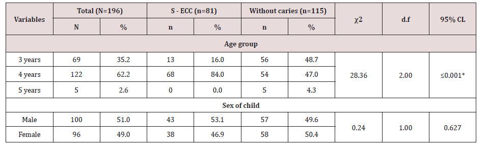
There were 196 children aged between 3-5-years-old who were recruited into the study, eighty-one children with S - ECC (41.3%) and 115(58.7%) without caries. The children’s mean age was 4.1+0.6 years, and it ranged between 3 and five years with a high proportion of the children (62.2%) aged four years. There was a statistically significant difference in age distribution among children with Severe. ECC and children without caries (χ2=28.36, d.f =2, p<0.001). The majority of the children with caries were aged four years (84.0%) compared to those without caries (47.0%). Gender distribution was comparable with boys slightly more (51.0%) than girls (49.0%) Table 1.
Table 1: Effect of group on prevalence of number of analgesic tablets at pre-treatment time interval.
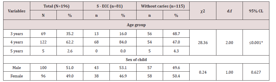
Lupinepublishers-openaccess-dentistry-oral-healthcare
*Pearson’s Chi-square
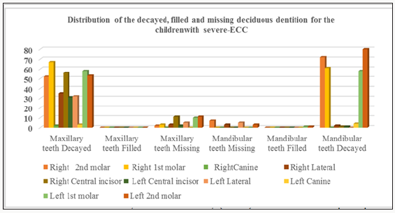
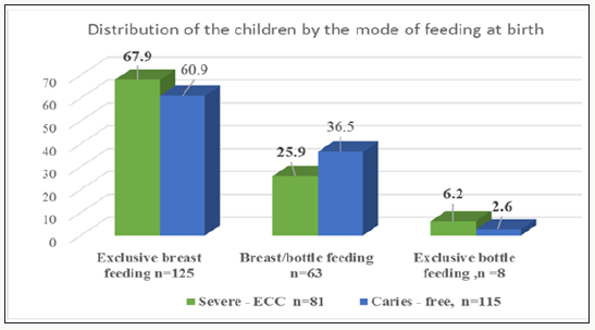
Out of 3,240 deciduous teeth for 81 children aged 3-5 years sixty one 1.9% of the teeth were missing due to decay hence 3179 teeth were examined of whom 605 (19%) were decayed... The caries pattern was that the maxillary teeth were more affected compared to the mandibular teeth. In the mandible the least affected were the canines. However the first primary molar had high prevalence of between 71.6% - 82.7%. In the mandible the most affected teeth were the second deciduous molars which had a prevalence range of 88.9% - 98.7% Figure 1 the study and they did not have ECC had been exclusively breastfeeding. However, 71 (36.2%) out of the 196 respondents had had both breast/bottle feeding. Out of the seventyone, those who had breastfeeding supplemented with bottle feeding were 29(35.8%), and they had severe ECC while 42(36.5%) out 115 of those without caries. Children who were exclusively bottle fed were eight of whom five 6.2% had severe ECC while three 2.6% did not have caries Figure 2. There were no differences in the methods of breastfeeding with a Pearson Chi-square =3.51, d.f= 2, p=0.173 at 95 % CL.
Figure 1: Prevalence of number of analgesic tablets in the two groups after 24hr.
Lupinepublishers-openaccess-dentistry-oral-healthcare
Figure 2: Percent distribution of children by the method of feeding in infancy and toddler stage.
Lupinepublishers-openaccess-dentistry-oral-healthcare
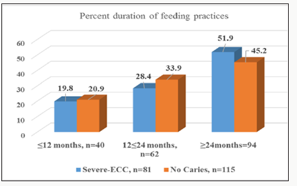
Forty children, 20.4% had breast or bottle feeding or combined feeding for a ≤12 month exclusively. Sixty-two (31.6%) 12≤ 24 months while 94(48%), for ≥24 months. There were 16 (19.8%) of the children with severe-ECC had either breastfed or bottle fed or both for a time duration of ≤12 months while those without decay were 24 (20.9%). Similarly, 23 (28.4) children with severe-ECC had a duration of 12≤ 24 months while those without caries were 39 (33.9%). Forty-two (51.9%) and 52(45.2%) of children with severe ECC and children without caries had respectively been breastfed for ≥24 months, Figure 3. However, there were no significant differences between the breastfeeding period for the children with severe –ECC and those without decay with a Pearson Chi-square = 0.92, d.f=2, p=0.630 at 95%CL.
Figure 3:Percent distribution of duration of feeding practices in infants and toddlers with severe-ECC and those without decay aged 3-5 years.
Lupinepublishers-openaccess-dentistry-oral-healthcare
The effect of amoxicillin with clavulanic acid antibiotic premedication on pretreatment pain after administration of antibiotic and before initiation of endodontic treatment (Pretreatment pain) was assessed for patients using a four step pain scale (No pain, Mild , Moderate, Severe). The results showed no statistically significant difference detected between both groups. The results are illustrated in the following images 3.
Figure 3: Effect of group on prevalence of pain score at pretreatment time interval.
Lupinepublishers-openaccess-dentistry-oral-healthcare
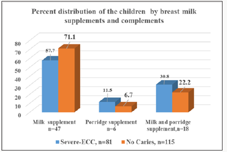
One hundred and twenty-five (63.8) children had exclusive breastfeeding while the remaining 71 (36.2%) had either breastfeeding supplemented with bottle feeding or exclusive bottle feeding. The seventy71 who had breastfeeding and supplement or had exclusive bottle feeding the breast milk complement or supplement used was either cow’s milk, porridge, milk mixed with porridge. Milk was the most common beverage bottle content for both groups of severe ECC and those without decay for 47 (66.2%) out of the sixty-three children who had been bottle fed; six 8.5%had porridge, For eighteen (25.4%) children the bottle content was a mixture of milk and porridge. Fifteen (18.5%) children with severe- ECC had milk as the bottle content while 3(3.7%) the content was porridge and eight ((9.9%) children the bottle content was milk and porridge Figure 4. There were no differences for the different practices about the breast milk complements or supplements with a Pearson Chi-square 1.39 d.f=2 p=0.500 at 95% CL.
Figure 4: Percent distribution of children of on breast milk supplements and complements using the bottle.
Lupinepublishers-openaccess-dentistry-oral-healthcare
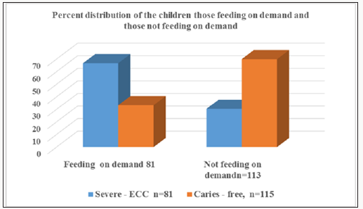
Eighty-nine (45.4%) children out of 196 were fed on demand while 107 (54.6%) were not fed on demand. Out of the 81 children with severe ECC 54(66.7%) were fed on demand compared to 35 (30.4%) out of 115 who did not have decay. However, 27 (33.3%) children out of 81 of those who had severe-ECC were not fed on demand. Similarly, 80 (69.6%) of the 115 who did not have decay were not fed on demand Figure 5. The difference was statistically significant with a Pearson Chi square= 25.17 d.f= 1.0, p≤0.001 at 95%CL.
Figure 5: Percent children of children who were fed on demand and those not feeding on demand for those with severe-ECC and those without decay.
Lupinepublishers-openaccess-dentistry-oral-healthcare
Six (3.1%) children had excellent oral hygiene, and they were from the group of children without decay. Children with good oral hygiene were 32 (16.3%) of which 10 (12.3) were from the group with severe-ECC and 22 (19.!%) from the group without dental decay.The oral hygiene of 148 (75.5%) children had affair oral hygiene, those with severe-ECC were 64 (79%) out of 81, and those without decay 84 (73%) out of 115 had fair oral hygiene. Only ten (5.17%) had poor oral hygiene of out of whom – (8.6%) had severe –ECC and three (2.6%) did not have decay, Figure 5. There was significant the difference in the oral hygiene status of children with S - ECC and children without caries with a Pearson Chi-square 2=9.18, df1, p=0.027).

Discussion

Severe early childhood caries (Severe–ECC) is an aggressive form of dental caries in the primary dentition associated with specific patterns of dietary intake in young children [1.10]. Most of the children 125(63.8%)were breastfed while 71(36.2%) were put on breastmilk compliments/ supplements early in infancy and some of them stayed on the bottle after the second birthday. Mothers in Kenya are encouraged to practice exclusive breastfeeding [8-14]. It is documented that nursing mothers in Kenya have a high breastfeeding frequency pattern occurring in 93% of mothers wherein a twenty-four hour period in the daytime the infant according to UNICEF a mother is recommended to breastfeed three times a day. However, Kenyan mothers are encouraged to breastfeed as much as possible and some of them breastfeed on demand as many as seven times in the daytime and five times at night on demand [11-13]. In the current study 63% of the children were exclusively breast fed and this finding is in agreement with the national value of 61% breastfeeding mothers who practice exclusive breastfeeding at least the first six moths of infancy. In order to enhance good oral health and the general health of and infant there is a need to provide information on the benefits of good oral hygiene for the breasting mother and the breastfed infant or toddler so that frquency of nocturnal at will breastfeedin is minimised . Thse may reduce the sustrate which the cariogenic bacteria require to produce acid and it will also reduce the production of the plaqure which holds the acid close to the enamel resulting in enamel dimineralisation. The oral health information and education may be incorporated in the prenatl clinics as information available to the expectantnt mothers.
It is currently documented that exclusive on demand breastfeedng may lead to severe- ECC which is a debilitating oral disease condition and it may be a confouder to malnutrition of the child who is in pain is unable to jew food properly and this may lead to nutritional deficiencis. The deficieneces may interfere with the proper pysical and mental growth and development of the child. Secondly vital nutional deficiences may lower the immunity of the child thus making the infant and toddler with severe –ECC to be vulnerable to early childood diseases.
The children who had exclusive breastfeeding were 63.7% of the study group and the breastfeeding period was ≥24 months which was slightly higher than the reported duration of exclusive breastfeeding [11,13]. Children who were fed on demand were 45.6% of the sample size out of whom 66.7% had severe ECC, and the difference was significant with a Pearson Chi-square p≤0.001.At will breastfeeding/ bottle-feeding on demand pauses a particular risk to the deciduous dentition which has low mineral content and thin enamel. In the current study out of 81 children with severe- ECC 55(67.9%). Out of the ninety-four children with prolonged breastfeeding 49(51.9%) had severe-ECC and had beyond twentyfour months. Though breastfeeding is good for the child, the nocturnal breastfeeding and the high frequency in the daytime which is twice what is recommended by UNICEF the stagnation of milk around the newly erupted teeth may be fermented by the anaerobic bacteria thus producing large quantities of acid. There is a need to encourage the mothers in breastfeeding but give them the knowledge to clean the infant’s mouth and to avoid nocturnal breastfeeding [11-13].
The dietary weaning practices included the use of a bottle where the breast milk supplement or complement was, milk, porridge or porridge mixed with milk. The introduction of a bottle has been associated with diarrheal disease in early childhood. The early childhood diseases further weakens the child’s immunity resulting in a vicious circle of disease and malnutrition in early childhood which may result in a child not being able to thrive.
Majority of the children with Severe -ECC were fed on demand (66.7%) compared to those without caries (30.4%), and this was statistically significant Pearson chi-square with p≤0.001. The difference in the oral hygiene status of children with Severe - ECC and children without caries were substantial with a Pearson Chisquare =2=9.18, df2 p=0.027).
Figure 6:Oral hygiene status for children with severe-ECC and those without caries.
Lupinepublishers-openaccess-dentistry-oral-healthcare
The most critical period of feeding at will has been reported to be twelve months.The period of twelve months is when most of the deciduous teeth with a thin and poorly mineraised enamel are fully erupted in the mouth. In the presence of the virulent anerobic bacteria Streptococcus Mutans Scardovia Wiggsiae the denttion is dimineralised [3,14]. The sustrate and bacteria presence are confounded by the factor that at one year there are no oral hygiene paracties for the toddler and the vist to the dentist is not yet hence emanel demineralisation may progress unabated Figure 6.
The feeding on demand results in having acid producing bacteria resulting in prolonged periods of a low pH resulting in the demineralization of the dentition. Recent research has incriminated the bacteria Scardovia wiggsiea as an anew pathogen which has been found at the sites of severe EC lesions in the absence of other pathogens which had previously been associated with severe ECC [2,3]. Ultimately, prolonged exposure of the teeth in the acidic environment causes dental caries. There were differences in the oral hygiene status of the children with severe –ECC compared with those who had no caries which was statistically significant.
A study in Saudi Arabia has reported similar findings where caries was associated with a high presence of debris [6]. The high caries debris could probably be due to poor oral hygiene practices among children with severe– ECC. National Oral health survey has reported poor utilisation of oral health services where a sample of 2,126 individuals age 5-15, nine hundred and only three (46.7%) had never visited an oral health facility. Out of those who had never visited a dentist, 57.7% were from the rural community where the services were scarce due to distance or resources were not available to provide oral health services for both the children and the adults [15,16].
The challenge may be overcome by having information and education incorporated in the well established maternal health, and well-child clinics on simple preventive remedied for good oral health practices to minimize plaque deposits and severe ECC.The preventative measures may ensure that the children have healthy teeth for mastication and digestive processes would also improve the quality of life for the children.

Read More Lupine Publishers Blogger Articles Please Click on: https://lupinepublishers.blogspot.com/
Read More Lupine Dentistry Journal Blogger Articles Please Click on:
https://lupine-dentistry-oral-health-care.blogspot.com/

Thursday, March 21, 2019

Determination of Alkaline Phosphatase Level in Gingival Crevicular Fluid during Rapid Maxillary Expansion(MADOHC) - Lupine Publishers

Determination of Alkaline Phosphatase Level in Gingival Crevicular Fluid during Rapid Maxillary Expansion(MADOHC) - Lupine Publishers


Introduction: Rapid maxillary expansion (RME) is a remarkable and ideal procedure which has a long history of over 100 years. When a transverse maxillary Arch is diagnosed it is treated by doing RME. Although numerous studies have been done in last four decades on gingival crevicular fluid, no data have been reported on the repeatability of gingival crevicular fluid collection and then following its quantification procedure. The objectives of the study were to observe the changes in alkaline phosphatase (ALP) activities in gingival crevicular fluid (GCF) during rapid maxillary expansion and particularly in retention period and then explore the relationship between these changes and periodontal tissue remodeling.

https://lupinepublishers.com/dental-and-oral-health-journal/fulltext/determination-of-alkaline-phosphatase-level-in-gingival-crevicular-fluid-during-rapid-maxillary-expansion.ID.000115.php

For more Lupine Publishers Open Access Journals Please visit our website: http://www.lupinepublishers.com/


Tuesday, March 12, 2019

Medical Leadership, Personal View (MADOHC) - Lupine Publishers

Medical Leadership, Personal View (MADOHC) - Lupine Publishers

Opinion: What exactly does "medical leadership" mean? To be honest, this made me ponder on that very question. For over a year I've been serving in the "Military Academy of Medicine" as a commander of a variety of courses - involving military trainings of dentists, dental assistants, nurses and more. I speak of the importance of being an officer in the medical corps of the IDF on a weekly basis and yet I came to the realization that I, myself, am not sure of its importance. The time I have spent thinking and planning before writing this essay has made me dig inside and eventually made me reach a better understanding of my role as a medical leader. Medical leadership can be divided into several parts.


For more Lupine Publishers Open Access Journals Please visit our website: http://www.lupinepublishers.com/

Tuesday, November 13, 2018

The Dental Service of the French Army in 1940 - Lupine Publishers


On December 1 1892, the Brouardel law was voted which gave legal status to dental surgeons. At that point, those who had followed appropriate studies delivered by the Faculty of Medicine were the only ones who could practise dental surgery [1]. Therefore, it was a profession in its infancy which enlisted during the war in 1939? On February 26 1916, a dental service was created in the infantry within the French army for the duration of the war. That of the Navy only started on March 1st 1916. After the Great War, only the Navy decided to dissolve its dental service for it did not have a satisfying organization to greet its patients. In 1934, a backup dental service was implemented once again. While they were made lieutenants in 1818, dentists could become captains after the law voted on December 19 1934 [2]. A few months later, dentists in the land army were attributed the same rank.  


For more Lupine Publishers Open Access Journals Please visit our website: http://www.lupinepublishers.com/
For more Dentistry and Oral Health Care articles Please Click
 Here:  https://www.lupinepublishers.com/dental-and-oral-health-journal/index.php


Lupine Publishers: Lupine Publishers | Post Endodontic Pain Reduction...

Lupine Publishers: Lupine Publishers | Post Endodontic Pain Reduction... : Lupine Publishers | Journal of Otolaryngology Research Impact Fac...