Showing posts with label Lupine Publishers. Show all posts

Sunday, March 1, 2020

Lupine Publishers | Oral Hygiene Habits and Dental Treatment Needs of Children with Dental Fluorosis and Those Without Dental Fluorosis Aged 12-15 Years In in a High Fluoride Area in North Kajiado Kenya

Lupine Publishers | Journal of Oral Healthcare

Abstract


Background: The dental disease identified as dental caries, periodontal, gingival lesions and dental fluorosis when diagnosed early and the treatment needs assessment with patients’ perception ensures the proper use of the physical facilitates, It also enhances planning for rational health resource allocation, utilization and personnel distribution so as to tackle the health problems in a holistic way.
Objective: The objective of the study was to determine the dental treatment needs among children aged 13-17 years affected by dental fluorosis and those not affected by dental fluorosis in Kajiado North District of Kenya
Materials and Methods: Study design this was a cross sectional comparative study of the dental treatment needs among two age matched population groups in primary school children. Sampling and Sample size. Stratified random sampling was used to select four primary schools out of the primary schools in North Kajiado. All children in the school with a full permanent dentin and whose parents had signed the consent form were recruited into the study. The study involved 248 children, 98(40%) males and 150(60%) females aged between 13 -17 years (mean age = 14.75±1.45) selected by simple random sampling from 9 schools in Kajiado North District which was purposively selected. They were all clinically examined under natural light for plaque and gingival scores using the Silness and Loe 1, Loe and Silness, dental caries was recrded using the decayed Missing Filedl teeth (DMFT), while gingivitis, periodontal disease and fluorosis using indices:- Silness and Loe 1, Loe and Silness, DMFT,CPITN and TFI.
Results: The treatment needs for gingivitis were similar, majority 218 (88%) children with fluorosis and 213 (86%) without required oral hygiene instructions and prophylaxis. There were 3(1.2%) children who had periodontitis in the group with dental fluorosis and required scaling and root planning. There were 50% children with caries in the fluorosis group who required one surface and 24.2% for two surface amalgam/composite restorations and for those without fluorosis, 76% required one surface and 15.2% two surface amalgam/composite restorations. There were 321(60.8%) teeth surfaces which required bleaching and microabrasion or composite masking and another 207(39.2%) for direct composite / porcelain veneers or crowns.
Conclusion: Children with dental fluorosis were burdened more by dental disease and had more treatment needs (dental caries, fluorosis, periodontal disease and gingivitis) when compared to those without dental fluorosis.

Introduction

Dental conditions like fluorosis, caries, gingival and periodontal diseases require varied treatment approaches to manage them depending on the severity hence the need to establish the levels of disease burden and treatment needs for proper planning of dental services. Well assessed dental treatment needs go a long way in the estimation of resources, rational fund allocation and efficient utilization of dental materials. Dental treatment needs should be assessed objectively and subjectively based not only on normative assessment but also on perceived needs and impact so as to obtain the best outcomes. The incorporation of both the clinician’s objective assessment and the client’s felt needs is essential in ensuring that they participate in the general management of their condition. In common practice today Bradshaw (1978) in a study on the problems and progress in medical care said that the treatment needs of most dental conditions are based on the clinician’s judgement using the recommended dental indices1. The age between 13-17 years forms the transition period between childhood and adulthood. The growth changes seen during this stage of life warrants a clear understanding of the health needs in general and support for optimal psychosocial and emotional development. Welbury pointed out that children affected by fluorosis suffer from low self - esteem, social stigma and poor performance in school.
Facial image is an important aspect with regard to an individual’s presentation and self-esteem in communication. This is greatly affected by the presence of dental fluorosis among other things like mal-aligned teeth, missing anterior teeth or even congenital malformations of the oral cavity. Globally there is often a permanent stigma associated with dental fluorosis among children or adults. A study conducted in Brazil by Rodriques showed esthetic changes in the permanent dentition are the greatest concern in dental fluorosis. Studies by Welbury and Glasser have observed that if left untreated, dental fluorosis causes embarrassment, psychosocial distress, difficulties in societal adjustment, damage to self-esteem and poor performance for the school-going children. Another study in Kenya by Mwaniki showed that between 60.4% and 84.3% of the respondents viewed dental fluorosis as a problem because of its unfavourable effects on an individual’s personality. It is important to note that dental fluorosis leads to shyness in expression thereby masking the true personality of an individual. It is further evidenced by a South African study by Mothusi that showed the trauma suffered by young people with dental fluorosis to be depressing such that they requested to have the teeth extracted and replaced with dentures.
Generally, the quality of life is greatly affected by oral diseases, dental fluorosis not being an exception, with a significant impact on the 13-17- year- olds due to their delicate stage of growth and development. Children experience appreciable impacts on oral health related quality of life with the greatest burden being associated with dental caries and to a lesser extent, fluorosis according to a study in Uganda by Robinson. The aspects considered when determining the quality of life with regard to oral diseases using the oral impact of daily performance (OIDP index) include eating, speaking and pronouncing clearly, cleaning teeth, sleeping and relaxing, smiling without embarrassment, maintaining emotional state and enjoying contact with others. A study in Tanzania by Roman on the impact and treatment needs of dental fluorosis where a total of 269 students with dental fluorosis aged 15-18 years (mean age 17.3) were involved, showed that a majority (65.4)% had severe dental fluorosis (TFI 6-9) while 29.4% had TFI 4-5 and 5.2% had TFI 1-3. Most of the students in this study (92.6%) perceived at least one (OIDP) with the most affected being smiling at 88.1%, emotional stability 81.4%, and having contact with others 75.5%. Studies by Locker and Leake indicated that the oral health status of at risk children and adolescents appeared to have been poor resulting in the need for several treatments including urgent, restorative, periodontal and preventive care Table 1.
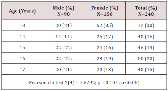
Table 1: Distribution of study participants according to age and gender.
Materials and Methods

Study Population

The study population comprised of 13 -17- years who were born and brought up in Kajiado North District in the first 7 years of life. The target population involved 34,122 children aged 13- 17 years according to the Kenyan population and housing census 2009 for Kajiado North District. The public primary and secondary school enrolment was approximately 19,065 for the ages 13-17 years in the year 2011 in the study involved 248 children, 98(40%) males and 150(60%) females aged between 13 -17 years (mean age = 14.75 ±1.45) selected by simple random sampling from 9 schools in Kajiado North District which was purposively selected. They were all clinically examined under natural light for plaque and gingival scores, dental caries, gingivitis, periodontal disease and fluorosis using indices:- Silness and Loe 1963, Loe and Silness 1964, DMFT,CPITN and TFI. Information on biodata, consumption of sugary snacks, brushing was collected using an interviewer administered questionnaire. Water samples were collected for testing for fluoride levels at the government chemist laboratories.

Data analysis

The clinical examination forms were pre-coded. The quality of data was ensured during the entire study process especially at the data collection point to include completeness of questionnaires, and validity of responses. Data was de - indentified and stored in a password protected data base with access being granted to the statistician. Quality control through data cleaning and validation was censured by counter checking frequencies in the computer and any missing data was re - entered. The findings from the study were organized in the form of frequency tables and figures. Computations to calculate disease burden (caries experience, prevalence of gingivitis and periodontitis, treatment needs and the cost of treatment) were done. The independent variable for this analysis was presence/absence of fluorosis while the dependent variables were age, gender, gingivitis, periodontitis, caries experience and cost of treatment. The confounding factors were snacking and oral hygiene practices. For categorical variables association between dependent variables and fluorosis was tested using a Pearson Chi-square test while a student t-test was used for continuous variables and the conventional P value of cut-off of < 0.05 was used to establish a significant association. To calculate the total DMFT, the total number of teeth per child with caries, filled due to caries, missing due to caries was summed up. For the mean gingival and plaque scores, the total score per child was calculated by summing the individual tooth scores, divided by 6 and the total for the index teeth added and divided by 6. To determine the agreement rates between assessors, a Cohen kappa score (agreement rate) was calculated for each assessment (tooth and surface) for all children assessed. A median agreement rate was then computed from all individual scores calculated. Data collected was analyzed using statistical package for social sciences (SPSS version 17.0) Table 2.
Table 2: Sources of drinking water.
Results

Socio demographic characteristics

This study involved 248 children aged between 13-17 years with a mean age of 14.75 years (±1.45 SD) who were all matched for age and gender. The ratio of children with dental fluorosis and those without was 1:1 and the male to female ratio was 2:3 and was not statistically significant [p= 0.104 (p ≤0.05)] as shown in Table 1. There were 241 (97%) participants born and raised in Kajiado North while 7(3%) moved to the district before 7 years of age.

Source of water and analysis

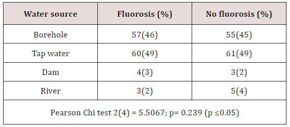
There was a similar pattern on the water sources which was not statistically significant [p=0.239 (p≤0.05)] for children with fluorosis and those without fluorosis. Most of the study participants consumed borehole water and most of tap water was also from boreholes. Dams and river sources were for a minority group as shown in Table 3.
Table 3:
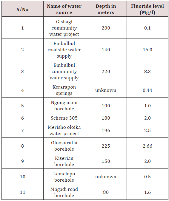
Gishagi borehole which is in a raised ground recorded low fluoride levels of 0.1 ppm as well as Kerarapon springs 0.44 and Lemelepo borehole 0.5, Ngong main borehole had the recommended levels by WHO of 1ppm. Embulbul roadside and Embulbul community water supplies had very high levels of fluoride at 8.3 and 15ppm (Table 4).
Table 4:
Tooth brushing habits

Majority of the children 122(98%) from each group brushed their teeth, the frequency of brushing was similar where by 113(93%) with fluorosis and 105(88%) without used a toothbrush while a chewing stick was used by a few (Table 5). The type of tooth brushing aid used was not statistically significant p=0.120(p≤ 0.05). Majority brushed once a day either in the morning after breakfast 61(50%) and 59(48%) or in the evening after meals 49(40%) and 51(42% for the children with fluorosis and those without fluorosis respectively. Only a small percentage brushed their teeth twice a day. There was no statistical significant difference on the timing of brushing between the groups [p=0.180(p≤ 0.05].
Table 5:
Relationship between brushing habits and plaque scores

Generally, children who brushed once after breakfast in both groups had PSs which were statistically significant p=0.003(p≤0.05) and the children with fluorosis had the lowest PSs of 0.85(0.5). The other brushing timings were not statistically significant p=1.02(p≤0.05) for at night and p= 0.664(p≤0.05) for twice a day as depicted in.

Gingivitis

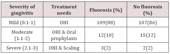
Both groups had a similar pattern of treatment needs. There were 109(88%) for OHI, 12(10%) OHI and oral prophylaxis, 3(2%) OHI and scaling for children with fluorosis. Those without fluorosis, 107(86%) OHI, 15(12%) OHI and oral prophylaxis, 2(2%) OHI and scaling.

Periodontitis

Of the 3(2.4%) children with fluorosis who had periodontitis they all required scaling and root planing.

Dental caries

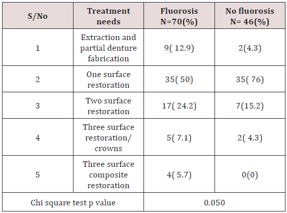
In both groups, dental restorations in form of one surface fillings were mostly indicated as 35(50%)/ 35(76%) for the children with fluorosis/those without. Two surface restorations 5(7.1%) for fluorosis and 7(15%) those without fluorosis. Extraction and partial dentures 9(12%) for fluorosis and 2(4.3%) those without fluorosis. Three surface composite restorations among children with fluorosis were 4(5.7%).

Caries experience in relation to consumption of sugary snacks

In the group with fluorosis the children who consumed sugary snacks twice had a higher DMFT of 0.71(1.4) while children without dental fluorosis and consumed four times scored highest DMFT of 0.83(0.9). On the weekly snack consumption, those who sacked once had no caries in both groups p=0;000(p≤0.05) which was statistically significant while the highest DMFT was recorded in those who snacked twice/four times for the fluorosis group at 0.64/0.6 while in the group without fluorosis the scores ranged between 0.17-0.25 despite different weekly snacking times.
None of the children who brushed twice had dental caries experience in the fluorosis group for once a day (after breakfast or at night) had a DMFT of 0.5(1)/ 0.73(1.6). For the group without fluorosis, there was some caries experience despite the timings for brushing. Generally there was no statistical significant difference on the brushing timing for both groups. The children who brushed after breakfast had a p=0.850(p≤0.05), at night only p=0.073(p≤0.05) and twice a day p=0.217(p≤0.05) therefore, brushing did not have any influence on the caries experience.

Cumulative TFI frequencies

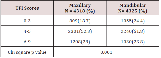
In both jaws TFI 4-5 was the most frequent at 2301(52.3%) on the labial and lingual surfaces of the anterior teeth in both the maxilla and of the mandibular anterior teeth 2240(51.8%). There were 321(60.8%) surfaces which required bleaching and/or micro abrasion or composite masking while 207(39.2%) surfaces required porcelain veneers or crowns Table 6.
Table 6:
Discussion

The current study did not find much difference in the treatment needs for gingivitis between the two groups as majority 88% with fluorosis and 86% without fluorosis required oral hygiene instructions and oral prophylaxis and in periodontitis the 1.2% affected required scaling and root planing. Most of the subjects with dental caries required some form of restorations either one, two or three surface amalgam/ composite restorations. A smaller number required extractions and partial dentures. Since radiographs were not taken for this study, it was difficult to ascertain the teeth which were indicated for pulp therapy. Studies done in Trinidad and Tobago by Naidu and Uganda by Nalweyiso clearly indicated that the treatment burden of dental caries is mainly centered on fillings, fissure sealants, pulp therapy and extractions. This study considered treatment needs for dental fluorosis in terms of labial surfaces from canine to canine in the maxillary teeth only. It was established that 48% of the teeth surfaces required bleaching / micro abrasion, composite masking and 52% for direct/indirect composite veneers/crowns. In Kenya Mwaniki found that 60.4 - 84.3% of the respondents viewed dental fluorosis as a problem although the study design was different from the current study.

Conclusion

Children with dental fluorosis were burdened more by dental disease and had more treatment needs (dental caries, fluorosis, periodontal disease and gingivitis) when compared to those without dental fluorosis.


Read more Lupine Publishers Blogger Articles please click on: https://lupinepublishers.blogspot.com/

Follow on Twitter       : https://twitter.com/lupine_online

Monday, February 24, 2020

Lupine Publishers | Down’s Syndrome- A Disease Caused By Genetic Alteration

Lupine Publishers | Dentistry Open Access Journal

Abstract

Down’s syndrome is the most common syndrome, medical professional encounters in day to day practice. It is a genetic disorder with a typical face profile and few classical intraoral features. Herein we report case and review on Down’s syndrome with facial features.
Keywords: Down’s Syndrome; Trisomy; Chromosome; Oral Manifestation

Introduction

Down syndrome is one of the commonest disorders with huge medical and social cost. DS is associated with number of phenotypes including congenital heart defects, leukemia, Alzheimer’s disease, Hirsch sprung disease etc. [1]. Down syndrome is a prevalent genetic disorder in intellectual disability in India. Its prevalence in tribal population is not known [2]. Down syndrome is one of the leading genetic causes of intellectual disability in the world. DS alone accounts 15-20% of ID population across the world [3,4].

Case Report

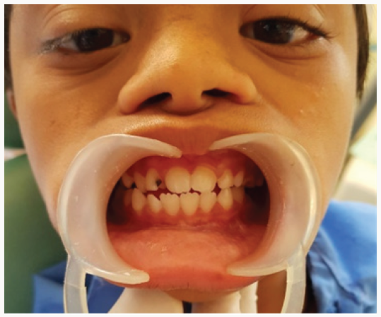
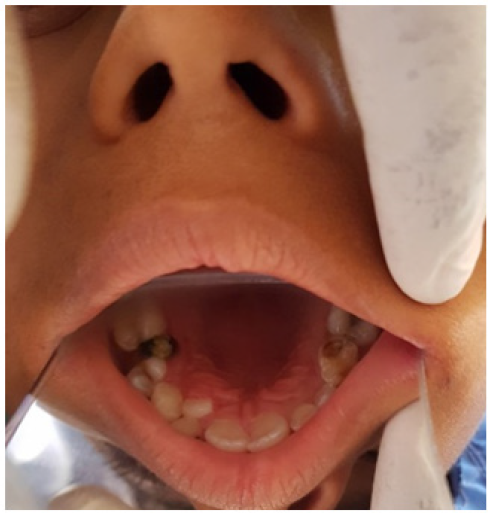
An 8 year old male patient came to the department of oral medicine and radiology for routine dental check-up. Extra oral examination revealed characteristic facial profile with increased inter canthal distance (Figure 1). Intraoral examination revealed Gingiva was soft with deposits on the teeth, High arched palate, with depressed nasal bridge was seen (Figure 2). Macro glossia was also seen .Correlating the intraoral and extra oral findings a Provisional diagnosis of Down’s syndrome/ Trisomy 21 was given. Patient was referred to the respective departments of pedodontics for restoration of decayed teeth.]
Figure 1: Extra oral features showing increased inter canthal distance and depressed nasal bridge.
Lupinepublishers-openaccess-dentistry-oral-healthcare
Figure 2: Intraoral features showing high arched palate.
Lupinepublishers-openaccess-dentistry-oral-healthcare

Discussion

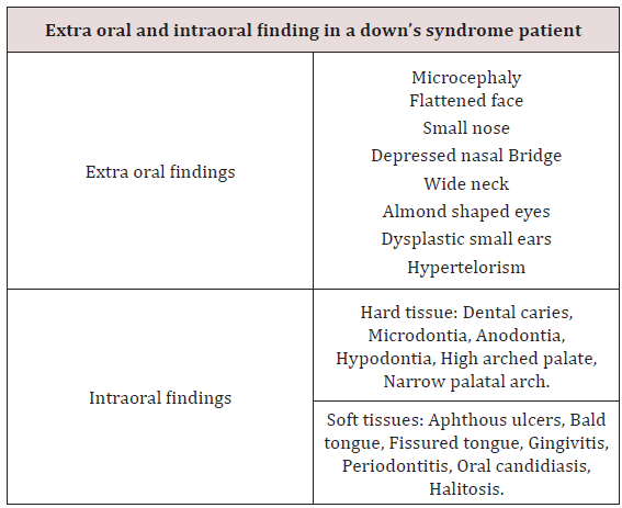
Down syndrome is one of the most leading causes of intellectual disability and millions of these patients face various health issues including learning and memory, congenital heart diseases, Alzheimer’s diseases, leukemia, cancers and Hirsch rung disease. The incidence of trisomy is influenced by maternal age and differs in population [5,6]. Facial findings in the patients can be characterised into extra oral and intraoral features (Table 1) [7]. Parents of children with Down’s syndrome should be aware of these possible conditions so they can be diagnosed and treated quickly and appropriately. According to Asim A et al. A Down’s syndrome child should have regular check-up from various consultants. These include:
a) Clinical geneticist - Referral to a genetic counselling program is highly desirable.
b) Developmental paediatrician.
c) Cardiologist - Early cardiologic evaluation is crucial for diagnosing and treating congenital heart defects, which occur in as many as 60% of these patients.
d) Paediatric pneumonologist -Recurrent respiratory tract infections are common in patients with DS.
e) Ophthalmologist.
f) Neurologist/Neurosurgeon - As many as 10% of patients with DS have epilepsy; therefore, neurologic evaluation may be needed.
g) Orthopaedic specialist.
h) Child psychiatrist - A child psychiatrist should lead liaison interventions, family therapies, and psychometric evaluations.
i) Physical and occupational therapist.
j) Speech-language pathologist.
k) Audiologist.
l) Paediatric dentist.
Hackshaw AK et al in their study, proposed a new screening method in which measurements obtained during 1st and 2nd trimester are integrated to provide the risk status of having pregnancy with DS. Moderate to severe intellectual disability occur as a constant feature, with IQ’s ranging from 20 to 85 [8]. Kennard in his review stated that there are a number of ultrasound markers in Down’s syndrome which includes nuchal fold thickness, cardiac abnormalities, duodenal atresia, femur length & pyelectasis [9]. The signs and symptoms of Down’s syndrome are characterised by neotenization of brain and bodies. Management strategies such as early childhood intervention, screening from common problems, medical treatment when indicated, a conductive family environment and vocational training can improve the overall development of children with Down’s syndrome [10].
Table 1:
Lupinepublishers-openaccess-dentistry-oral-healthcare

Conclusion

Genetics have always have played a major role in physical and mental being of an individual. Downs patients being mentally and medically weak, best care needs to be taken with adequate precautions.

Read more Lupine Publishers Blogger Articles please click on: https://lupinepublishers.blogspot.com/


Follow on Twitter       : https://twitter.com/lupine_online

Thursday, February 13, 2020

Lupine Publishers | The Nutritional Status of the Children with Severe- ECC Comparison with the Nutritional Status of Children without Caries Aged 3-5-Years-Old and with the Caregiver’s Demographics in a Kenyan Hospital

Lupine Publishers | Dentistry Open Access Journals

Abstract

Severe early childhood caries (Severe-ECC) is an aggressive, infectious and preventable form of dental caries that affects very young children. The survey purposed to examine any differences in the severity of poor nutrition in children without decay and those children with dental decay in the age group between thirty-six and sixty months. Sampling was purposeful and 196 children aged between 3 to 5 years for this study. The study was hospital-based where eighty-one children with severe dental decay who had attended the Nyanza Provincial General Hospital (NNPGH). Similarly, one hundred and fifteen children who were caries free were chosen from amongst the children attending the maternal child health clinic at NNPGH over a period of three months. Odds Ratio (OR) and 95% Confidence Interval (CI) were used to estimate the strength of association between Severe ECC and nutritional status. The mean dmft for the children with severe Early Childhood Caries (ECC) was 7.5±19. The prevalence of malnutrition was reported among both groups of children with severe ECC and without decay as 28 (14.3%) underweight, wasting 5(2.5%), and stunting 9(4.6%). The malnutrition in children with, Severe-ECC was observed as 27(14%) underweight; 10(4.9%) of the children were wasted, and 5(2.5%) were stunted. However among the children without caries 26 (13.9%) were underweight while 5 (2.6% were wasted, and 12 (6.1%) were stunted. Both children those with severe ECC and those with decay, however, the children who were likely to be underweight at 1.23 times were those affected with severe ECC at 1.23 times compared to the children without decay. Hence other factors may be playing a role in malnutrition of children aged 3-5year old.
Keywords: Severe-ECC; Nutritional status; Caregivers demographics

Introduction

Early childhood caries (ECC) is defined as the presence of one or more decayed (non-cavitated or cavitated lesions), those missing (due to caries), or filled tooth surfaces in any primary tooth in a child 71 months of age or younger. Severe Early Childhood Caries reported in children below three years of age as smooth surface caries1. One or more cavitated, missing teeth due to caries has been associated with age s 3-5years.The filled smooth surfaces in primary maxillary anterior teeth or a decayed, missing or filled a score ≥ 4 for age 3years, a score of ≥ five is associated with 4years while cavitation, restored tooth and missing due to caries a score of ≥6 is for children in the 5-year-old group. All these scores constitute Severe – ECC [1].
Disadvantaged groups have been found to be vulnerable to ECC in both developed and developing countries and even within a single country disparity by social standing there exist, differences due to diet, fluoride use, and social empowerment. Disparities in social empowerment may persist due to lack of access to dental care and inadequate utilisation of dental care even when available [2]. Untreated caries and associated infections can cause pain, discomfort, reduced intake of foods because eating is painful
[3]. Pain may also because the child refuses the caregiver from maintaining good oral hygiene for the child. There is a paucity of literature on the prevalence of Severe -ECC in Kenya. However, a study conducted in nursery school children in Nairobi on the on dental caries and dietary patterns reported a prevalence of 63.5% among 3-5 years old [4]. A survey conducted in Kiambaa division in Kiambu County, a peri-urban population, reported ECC prevalence in 3 - 5-year-olds of 59.5% [5]. Several studies on nutritional status and dental caries have reported variable results. A retrospective survey on the body mass index was done in the United States of America, and it involved two hundred and ninety-three children aged two to five years with Severe - ECC receiving dental treatment under general anaesthesia. In the study, the weight groups were defined by being assigned the CDC body mass index about on age and gender of the children. Results showed that the distribution of subjects by percentiles and the children who were underweight were 11%; of the study sample. However the children whose weight was normal weight 67%; at risk of overweight 9%; overweight 11%. This study concluded that significantly, more children in the sample were underweight than in the reference population [6]. However comparative research on the nutritional status and dental caries among a large sample of four and five-year-old South African children found no significant association between the prevalence of caries and stunting or wasting. However, a relationship was found between decayed, missing and filled surfaces and wasting [7]. This study, therefore, aimed to compare the nutritional status of children aged 3 – 5 years with Severe-ECC and the nutritional status of those aged 3-5 years without caries.
Severe ECC is also associated with oral Microbiota, and in particular anaerobic bacteria of the species Scardovia Wigggsiae and others have been found in abundance in severe ECC lesions [8]

Materials and Methods

One hundred and ninety-six children aged between 3 to 5 years were recruited for this study. Purposive sampling was done to select Eighty-one children with Severe - ECC was chosen from amongst the patients who had sought dental treatment at the dental clinic at the Nyanza Provincial General Hospital (NNPGH). However, 115 children who were caries free were selected from amongst the children attending the maternal child health clinic at NNPGH over a period of three months. Inclusion criteria were: the child was 3 – 5 years of age, was medically healthy, and the parent or caregiver was willing to consent. A semi-structured questionnaire was administered to the caregiver in a face to face interview, and information was collected on the socio-demographic background of the children. There gathered data included education level, age, gender, and the caregiver’s, occupation, and area of residence of the caregivers. The Intraoral examination was carried using dental mirrors and a Michigan O dental probe under natural light as the child sat on an ordinary chair facing the light. Severe ECC was defined as decayed, missing or filled a score of ≥ 4 (age 3), ≥ 5 (age 4), ≥ 6 (age 5). Before dental caries diagnosis, each tooth was dried using a piece of sterile gauze. WHO 1997 caries diagnosis criteria were used, and dental caries was diagnosed when there was a clinically detectable loss of tooth substance and when such damage had been treated with fillings or extraction [9]. Anthropometric measurements were determined to assess the nutritional status of the children and height of the children were obtained by measuring the child standing when standing erect and barefoot, using a measured with a standard height board to the nearest 0.5cm. Weight for age was measured using a Salter scale to the nearest 0.1kg. Each parameter of height and weight had three measurements taken, and an average of each was then recorded. The Cut-offs +2 standard deviations (SD) were used to identify children at significant risk for either delayed (<-2SD) or excessive (>+2SD) growth. The indicators were weight-for-age (WAZ), height-for-weight (HAZ), weight-forheight (WHZ) based on the World Health Organisation(WHO) 2005 recommended reference standard [10]. The collected data collected were coded, cleaned and analysed using SPSS version 17.0 (SPSS Inc, Chicago Illinois, USA) for Windows and Microsoft Office Excel 2007. Nutritional data was analyzed using Epi-Nutri program of Epi-Info version 3.5.1. Descriptive statistics such as proportions were used to summarize categorical variables while measures of central tendency such as mean, standard deviations and ranges were used to summarise continuous variables. The strength of association was established between categorical values using a Pearson’s Chi-square tests. Odds Ratio (OR) and 95% Confidence Interval (CI) were used to estimate the strength of association between independent variables and the dependent variable. The multivariate analysis was done using binary logistic regression at a statistical significance set at p≤0.05. The relevant research and ethics approving institutions approved the study.

Results

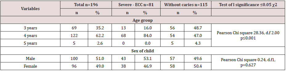
A total of 196 children aged 3-5 years were recruited into the study, eighty-one children with S - ECC (41.3%) and 115(58.7%) without caries. The study group had a mean age of 4.1 + 0.6years, and it ranged from 3- 5 years with a high proportion of the children (62.2%) aged four years. There was a statistically significant difference in age distribution among children with Severe ECC and children without caries (χ2=28.36, d.f=2, p<0.001). The majority of the children with caries were aged four years (84.0%) compared to those without caries (47.0%).Gender distribution was comparable with boys slightly more (51.0%) than girls (49.0%).
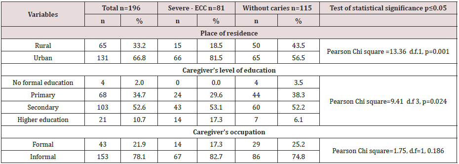
Sixty-five children (33.2%) lived in the rural community, and 131(66.8%) lived in the urban area. The differences in the area of residence were significant with a Pearson chi square=13.36, df=1, p≤0.001) for the children with severe ECC and those children without decay. It was noted that sixty-six (81.5%) out of 81 children with Severe ECC lived in an urban setting when compared to children who were caries- free who had 65 (56.5%) out of 115 children who were caries free. Some sixty-eight caregivers had had primary school education of whom 24 (29.6% had severe ECC while 44 (38.3%)) were caries free. However, 103 caregivers had secondary school education of whom 43 (53.1% had severe-ECC and60 (52.2%), while 21 (10.7%) their caregivers had tertiary education and 14 (17.3%) and seven 6.1% were caries free. Also, children whose caregivers had a primary level of education had the highest prevalence of severe-ECC followed by those whose caregivers had secondary education. The differences in the severecares prevalence were significant with a Pearson Chi-square =9.41 d.f 3, p≤0.024 Table 1 & 2.
Table 1: Age and gender distribution of children with Severe - ECC and children without caries.
Table 2: Level of education, demographics for the caregivers, place of residence, level of education, and occupation.
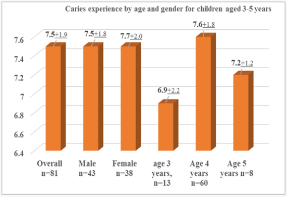
The mean dmft of 7; 5±1.9 d was observed among children with Severe – ECC, and it ranged from 5 to 12 scores. Scores. However, the mean dmft for the males was 7.5±1.8 and for females (7.5±2.0), which was statistically insignificant difference found between the two groups (t=0.15, p=0.88). The mean dmft score for children aged three years was 6.9 ± 2.2, four years was 7.6 ± 1.9, and for five-year-olds was 7.2 ±1.2 and all the dmft ranged from 5-12. The dmft progressively increased with age and peaked at age four years. There were no statistically significant differences found between the age groups (t=1.59, p=0.248). Figure 1.
Figure 1: Distribution of decayed, missing, and filled teeth by age and gender.
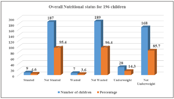
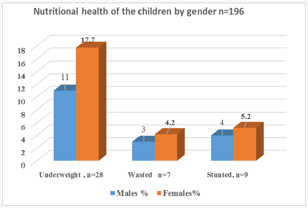
Overall the decayed component of the dmft contributed 92.3%. The missing and filled component of the dmft contributed 7.4% and 0.3% respectively. The overall prevalence of underweight for acute malnutrition, stunting, and wasting for chronic malnutrition was 14.3%, 4.6%, and 3.6% respectively. There were more females 17(17.7%), 4 (4.2%), and 5 (5.2%) who were underweight, wasted and stunted respectively when compared to males, but this difference was not statistically significant Pearson Chi-square respectively for underweight, stunted and wasted were 1.80,df=1, p=0.180 ; 0.19,d. f=1, p=0.660 and 0.16, d.f=1, p=0.686 Figures 2 & 3.
Figure 2: Prevalence of malnutrition for children aged 3-5 n=196.
Figure 3: Nutritional status by gender distribution.
Table 3: Underweight among children with caregivers place residence, level of education, and occupation.
When the caregiver’s residence, level of education, and occupation were considered the children who lived in the rural areas had higher prevalences of were underweight 10(15.4%), when compared to the children in the urban areas 18(13.7%) resided in urban areas. Sixty-eight children had caregivers whose education was of a primary level, and 11(16.2%) of the children were underweight while 57 (83.8%) had normal weight for age. Children whose parents had a secondary education were 103 of whom 14 (13.6% were underweight, and 89 (86,4%) had normal weight for an age while caregivers who had higher education were eighteen of whom 3(14.3%) were underweight, and 15( 85.7%) had normal weight. There were more underweight children 24(15,7) out of 153 when weight for age was examined about the caregivers who were informally employed, However, the differences in the children who were underweight with the caregiver’s various demographics were not significant Table 3.
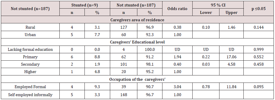
According to the educational level, the children who were stunted and whose parents had a primary education were four (9.3%)), secondary 6(8.8%), and higher education were 5(7.7%). The caregivers who had formally employed were from the urban area while those who were informally employed and had primary school education were from the rural areas Table 4. There were statistically insignificant differences in the caregiver’s place of residence, the level of education, and occupation among children who stunted and those who were not stunted.
Table 4: Stunting among children about caregivers place of residence, level of education, and occupation.
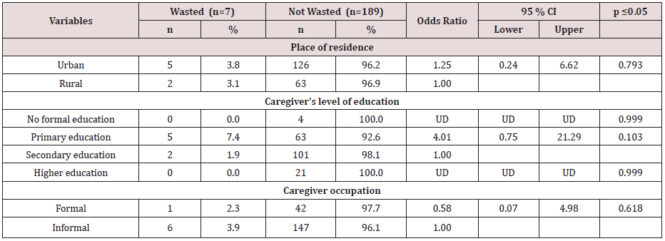
For the children who were wasted five 7.4% of the caregivers lived in the Urban area and had a primary level of education; also 6(3.9%) of the caregivers had informal employment, and 2(3.1%) resided in rural areas Table 5. There statistically insignificant differences in the caregiver’s place of residence, the level of education, and occupation among children who wasted and those who were not wasted.
Table 5: Wasting among children about caregivers place of residence, level of education, and occupation.
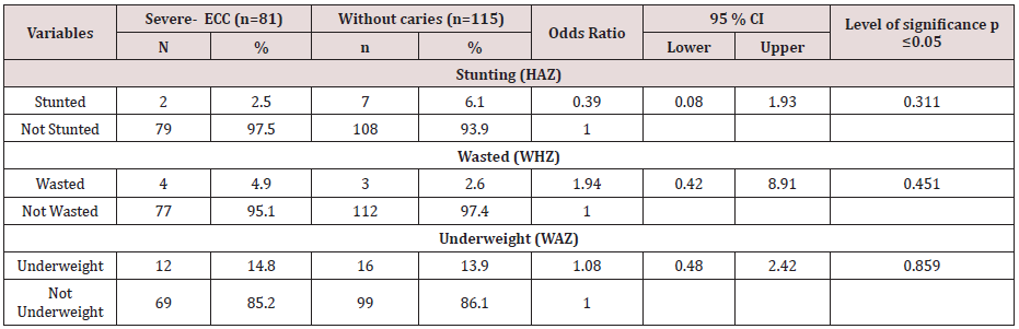
There was a slightly higher prevalence of underweight 14’8% for the children suffering from severe ECC compared with children without decay 13.9%. Although there were differences in the nutritional status of children with severe- ECC and children without caries the differences were insignificant for stunting with p=0.311; also underweight was insignificant with p=0.859 while wasting had p=0.451). A child identified with Severe- ECC at risk 1.08 more times likely to become underweight when compared to a child who did not have decay odds ratio lower and upper limits of 0.48 and 2.4 at 95% CL Table 6.
Table 6: Comparison of the nutritional status of children with Severe ECC and children without caries.
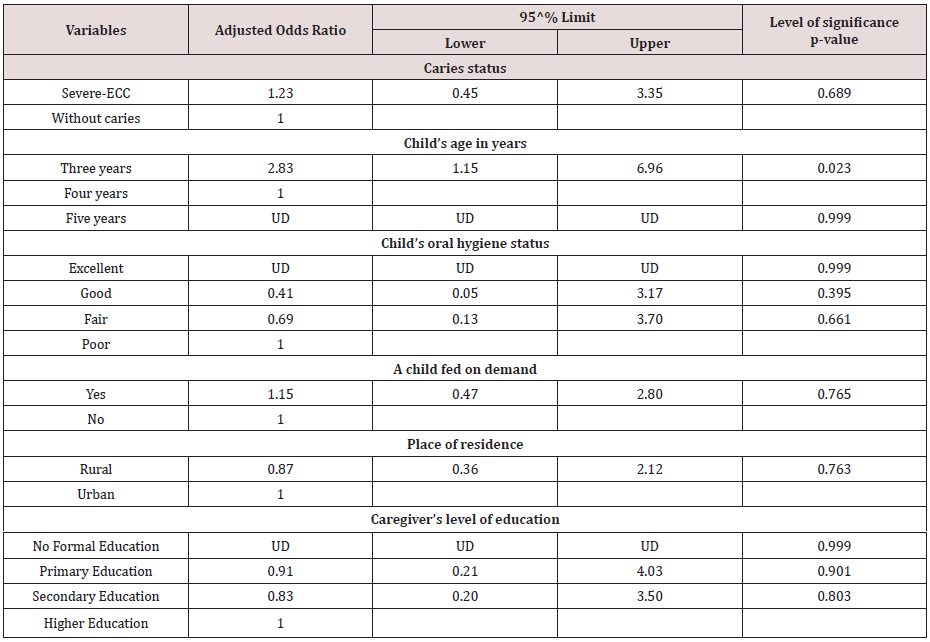
Multivariate analysis was done to determine the relationship between underweight and Severe- ECC among the participating children. Five factors associated with underweight and Severe- ECC at P≤0.05 during bivariate analysis were considered for multivariable analysis upon fitting the factors using binary logistic regression. Adjusting for child’s age in years, child’s oral hygiene status, child feeding on demand, place of residence and caregiver’s level of education, the occurrence of S-ECC was not significantly associated with underweight (AOR=1.23; 95% CI: 0.45 – 3.35; p=0.689). However, a child with S – ECC was 1.23 times more likely to have low weight for an age when compared to a child who was caries – free. However adjusting for other factors, age three years was found to be statistically significantly associated with underweight with an Adjusted Odds Ratio value =2.83; 95% CI: 1.15 – 6.96; p=0.023 Table 7. A child aged three years was 2.83 times more likely to be underweight when compared to one aged four years.
Table 7: Logistic Regression Predicting underweight using caries status, Child’s age in years, Child’s oral hygiene status, Child feeding on demand, Place of residence and Caregivers level of education.

Discussion

In the current study found that children with severe ECC were mainly from urban areas in comparison to children who were caries free. The finding of a high prevalence of severe –ECC in the urban children is similar to other studies in Kenya and elsewhere that have shown that children residing in urban areas have a higher caries experience than their rural counterparts [4,5,11,12]. The mean dmft of children with severe ECC in the present study was 7.5+1.9 which is comparable to a study carried out among preschool children of low socioeconomic status in India which reported a mean dmft of 8.9 [13]. Studies in the USA, and Canada among preschool children found mean dmft scores of 9.6±3.6 and 10.5 respectively [13-15]. The differences in the mean dmft may be due to variations in dietary practices among different populations. Also, decayed component accounted for 92.3% of the dmft, and this finding was similar to a study in South Africa [14]. Untreated tooth decay reflects a low utilisation of oral health services or lack and inaccessibility of preventive and curative dental services to the caregivers, or if the facilities are available, they are too costly.

Higher caries experience was observed in the children from the urban areas when compared to their rural counterparts [11]. The mean dmft of children with severe ECC in the present study was 7.5+1.9. The caries experience for severe-ECC in the present study is comparable to a study carried out in a low social, economic status in India among preschooler and reported a mean dmft of 8.9[112]. Studies in the USA, and Canada among preschool children have reported mean dmft scores of 9.6±3.6 and 10.5 respectively [13,14]. The differences in the dmft could be due to variations in dietary practices among different populations. The decayed component in the current study accounted for 92.3% of the dmft, which similar to other studies elsewhere [14]. Untreated tooth decay reflects a low availability and accessibility of preventive and curative dental services.

In this study, there were more females were underweight, stunted, and wasted when compared to males when referenced on the WHO reference standard. However, the differences were insignificant. The WHO child growth standards reference was used to evaluate nutritional status. The WHO growth reference provides a scientifically reliable yardstick of children’s growth achieved under desirable health and nutritional conditions and establishes the breastfed infant has been used as a reference against whom other alternative feeding practices are measured to and compare to regarding growth, health, and development of in children [9]. The children with severe-ECC who were underweight were 4.9%, stunted 2.5%, and those who were wasted were 14.8%. The presence of underweight, stunting, and wasting may be associated with the inability of the children with severe-ECC to chew the available food and absorb enough nutrients resulting in faltering nutritional status. In comparison a study carried out in Italy among 2- 6 years old found that 11% were e underweight, 11.11% overweight and 22.2% to be at risk of overweight [15]. A study in the USA reporting on the BMI of children with severe ECC noted those who were underweight as 11.%, overweight 11%, and those who were at risk of overweight were nine %6. These findings were insignificant may be due to differences in cultural, dietary practices and the primary determinants of nutritional status among the different populations. In Kenya, the primary determinants of nutritional status among children under five years of age include poverty, hunger, and drought [16]. The low weight for age observed with urban children is similar to previous research from other countries where children with high prevalence with severe-ECC had low weight for age [17].

Children who were malnourished were also noted to have severe ECC compared to children who were caries free. There are high levels of malnutrition in Nyanza as reported in the Kenya Demographic and Health Survey 2008-2009 where 19%, 2%, and 14%of the children under five years were underweight, wasted and stunted respectively [18]. Considering the caregiver’s demographic factors children who had low weight for age, wasting and stunted, resided in rural areas. Also, their caregivers had informal employment and had a primary level of education.The finding may be related to the low socioeconomic status and affect access to health care, food security and hence changing overall nutritional status [16,17].

The differences in the nutritional status of the children with ECC and those without ECC was insignificant. South African children aged between four and five years reported similar findings as what has been observed in this study. Njoroge et al. reported 60% in a study population of 338 children aged five years and below[4]. The most affected dentition were the upper central incisors however the severity of decay increased with age and the first and the second deciduous molars had the highest prevalence ranging between 57% -66%. In this study, the caregivers knew the importance of good oral hygiene and significance of snacks about caries formation. However, the infant feeding habits and the weaning practices were not reported on in this study [19,20].

The South African Study found no relationship between the prevalence stunting or wasting with dental caries. However, they reported an association between Wasting with the decayed, missing and filled tooth surfaces [7]. Children with severe ECC were 1.23 times more likely to be underweight when compared to children without caries. Severe ECC may affect general health and development because a toothache associated with caries may affect food intake and sleep [1]. Poor oral health associated with pain may interfere with the intake, mastication digestion of food and nutrients which may lead to decrease in good nutritional health and reduced quality of life for a child [1].

In summary, the difference in the nutritional status of children with severe ECC and children without caries and stunting was insignificant p=0.311, Underweight p=0.859 and wasting p=0.451. However, children with Severe ECC were 1.23 .times more likely to be underweight than children without caries.

Read more Lupine Publishers Blogger Articles please click on: 
https://lupinepublishers.blogspot.com/

Follow on Twitter       : https://twitter.com/lupine_online

Thursday, February 6, 2020

Lupine Publishers | Caregiver’s Oral Healthcare Practices And The Level of Utilisation Of Oral Health Services and The Dental Caries Experience Of 3-12-Year-Olds Suffering From Heart Disease in Nairobi, Kenya

Lupine Publishers | Dental and Oral Health Journals

Abstract

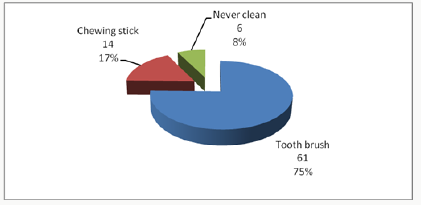
Cardiac diseases require that there is the meticulous maintenance of oral hygiene to avoid bacteremia, which has been associated with rheumatic heart disease and bacterial endocarditis. The aim was to establish the utilisation of oral health care and oral health practices of the caregiver about the oral hygiene and caries experience of children aged 3-12 years suffering from heart disease and were attending three pediatric cardiology clinics in Nairobi, Kenya. The study was descriptive and cross-sectional. It involved a study sample of children suffering from different types of cardiac conditions and attending the Pediatric cardiac clinics in three public institutions in Nairobi Kenya. The instruments the caregivers used to brush the children’s teeth were the toothbrush 61(75%); chewing stick 14(17%) and 6 (8%) never cleaned their teeth. Children who used a chewing stick had a lower dmft of 1.40±2.98 compared to a dmft of 3.22±3.59 among children who used the toothbrush, with Mann Whitney U, Z p=0.024 (p≤0.05).The children who brushed their teeth had a lower mean plaque score of 1.68±0.58 compared those who did not clean with a mean plaque of 2.28±0.40 with a Mann Whitney U, Z=-2.611, p=0.009(p≤0.05). It was noted that the children who had visited a dentist had a higher caries experience with a dmft of 4.18±4.13 and DMFT of 1.16±1.92. However, the children who had never sought treatment at a dental facility had lower dmft of 1.89±2.88; and DMFT of 0.36±1, and the differences were statistically significant with Mann Whitney U, Z p=0.008(p≤0.05). The plaque scores and caries experience were high in children whose caregivers had low aggregate utilisation of the oral health care facilities. However, those who had a low aggregate of oral hygiene practices had slightly higher plaque scores and caries experience.
Keywords: Cardiac Disease; Children; Utilisation; Oral Health Services; Caregivers

Introduction

Populations with chronic medical illness or other disabilities had the most unmet needs for oral health services [1], with poor oral hygiene and increased caries experience than the general population. For a child from a low-income family with heart disease, this means an added economic burden in an already tricky situation [2], as heart diseases necessitate regular dental check-ups and maintenance of meticulous oral hygiene. This concern has even been highlighted with new proposals on changes in the guidelines relating to prophylaxis against infective endocarditis [3,4]. The oral conditions may have a considerable impact on the general health status and quality of life of otherwise healthy children, but their effects on those children with acute and chronic illness can be more dangerous [5]. Children with cardiac defects and diseases are at increased risk or even life-threatening complications [6]. Hence the need for preventive dental health care geared to reducing the risks associated with management of the oral conditions under general anaesthesia. Also, the prolonged bleeding from warfarin medication often taken By the children [7-10]. Poor oral hygiene may give rise to a frequent bacteraemia under normal physiological conditions, and this can lead to a permanent risk of developing heart disease [11-14]. Two common oral diseases, namely periodontal and dental caries, though preventable, are still more prevalent in Kenya [15,16]. The children with heart disease have the disadvantage that their caregivers are preoccupied with the with the primary medical condition the cardiac disease, resulting in the neglect of other facets of the child’s total health [17]. The Kenya National Oral Health policy document has already indicated that the dmft value for Kenyan 5-year old children as at 2002 was 1.5±2.2, while 43% of 6-8-year-old children had caries [15], underscoring the fact that caries is still very rampant amongst the child population in Kenya.
The study was descriptive and cross-sectional where all the patients aged 3 to 12 years and their caregivers attending paediatric cardiology clinics over a three month period at Kenyatta National Hospital (KNH), Gertrude’s Garden Children’s Hospital (GGCH) and Mater Hospital. A Purposive sampling had been used to select the study hospitals. Based on Kliegman. study, the study population sample was determined as 79 cases. However, 81 patients were recruited in the study. A semi-structured questionnaire was used to collect information on the socio-demographic characteristics of the children and the parent/guardian habits on oral health practices and utilization of oral health services. As children waited to consult the cardiologist clinical examinations done to record the oral health status. The examination was conducted using sterilized instruments and under natural daylight, with the participants seated on a chair facing the window. Great care was taken during periodontal probing for gingivitis, to avoid initiating bleeding that could lead to septicaemia as the children were not on prophylactic antibiotics. The results were recorded on predesigned individual questionnaire sheets, and a record of dental caries and plaque was done. The dental caries was then recorded as dmft for the primary dentition and DMFT in the permanent [18,19], and the dental plaque was marked based on the Loe and Silness plaque score index [20]. Before commencement of the study, the examiner was calibrated by an experienced paediatric dentist on the collection of data relating to dental caries, and dental plaque Cohen’s kappa index score of 0.87 and 0.85 (n=10) was obtained for dental caries and plaque score respectively. The questionnaire was pre-tested before use. A duplicate clinical examination was also performed by the examiner to determine intra-examiner consistency, with results of Cohen’s kappa index score of 0.91 and 0.86 (n=12) being obtained for dental caries and plaque score respectively.

Data analysis

The data collected was cleaned, coded and analyzed using SPSS version 17-computer software from SPSS Inc. IL. The results obtained were compared and tested using Kruskal Wallis Chi-square and Mann Whitney U statistical tests, with statistical significance pegged at 95% confidence interval.

Results

The 81 children in the study, 44 (54.3%) were males and 37 (45.7%) females. Their ages ranged between 3-12 years with a mean age of 8.16 years (± 2.81 SD), and the 6-9-year-olds accounted for the most substantial proportion of 33 (40.7%) compared to the 3-5 year-olds who formed 16(19.8%). The differences in ages and gender were not statistically significant Chi χ2 =1.287, two df, p=0.525(p≤0.05). A total of 37(46%) children were from rural areas, 28(34%) were from Nairobi, and 16(20%) were from other urban centres other than Nairobi. The distribution of the children according to the type of heart disease, rheumatic (RHD) accounted for 36(44.5%) while infective endocarditis (IE) affected 4(4.9%). The duration since diagnosis of the cardiopathy ranged from less than one year to 12 years. Nearly half of the children, 40 (49%) had been diagnosed with the disease for a duration of between 1 to 5 years, while those who had been diagnosed more than five years and those less than one year accounted for 30% and 21% respectively. The caregivers’ oral health care practices that included how the child’s teeth were brushed; the frequency of brushing; and whether tooth brushing was supervised showed that 75(93%) children cleaned their teeth and 6(7%) children did not clean their teeth. Of the group that cleaned their teeth, 33(44%) did it twice a day, 29(39%) once a day while 16% once in a while/occasionally. About supervision, 62 (83%) reported cleaning their teeth without supervision while 13 were assisted by the caregivers. Inquiry on the ways the child’s teeth were cleaned, 75% (61) of the children used toothbrush and the rest of the results were as shown in Figure 1. The children who used toothpaste were 59 (79%) while 16 (21%) never use any toothpaste.
Figure 1:
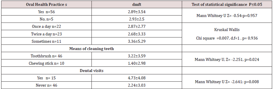
Considering the utilisation of oral health care services by children with heart diseases; fifty-nine (72.8%), children had never visited a dentist or utilised oral health services. Among the 22 (27.2%) children who had been to a dentist, the dental procedure during the last appointment included extraction 10 (12.3%). Also cleaning/prophylaxis (1(1.2%)), consultation ; check-up 9(11.1%) and fillings 2(2.5%).Caregiver’s oral healthcare practices and the dental caries experience about the children five children who never cleaned their teeth had a higher dmft of 2.93±2.50 compared to a lower dmft of 2.89 ±3.54 among the 56 children who cleaned their teeth, and the differences were insignificant with p=0.957(p≤0.05).
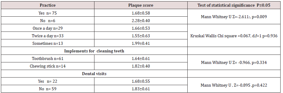
The differences in the frequency of tooth cleaning, the eleven children who cleaned their teeth once in a while had a higher dmft of 3.36±5.29 and the 23 children who cleaned twice a day had lower dmft of 2.68±2.77, but.difference was not statistically significant with p=0.936(p≤0.05). The children who used a chewing stick had a lower dmft of 1.40±2.98 compared to a dmft of 3.22±3.59 among the 46 children who used the toothbrush, with the difference was not statistically significant, p=0.024(p≤0.05). The children who had visited the dentist apparently had a higher caries experience with dmft of 4.18±4.13 and DMFT of 1.16±1.92 when related to the children who had never visited a dentist, who had lower dmft of 1.89±2.88; and DMFT of 0.36±1. These differences in the results were statistically significant, p=0.008(p≤0.05). The rest of the results are as shown in Table 1. When the caregivers were classified into two groups based on the responses to the oral healthcare practices as being favourable or unfavourable practices,53 (86%) caregivers fell in the unfavourable oral healthcare practices. Fiftythree children whose caregivers displayed unfavourable practices had a higher dmft of 3.62±3.54 compared to dmft of 2.74±2.85 among the eight children whose caregivers displayed favourable oral healthcare practices. The difference was statistically significant with Mann Whitney U, Z= -1.297, p=0.197(p≤0.05). The mean plaque score was significantly lower among the 75 children who reported to cleaning their teeth with mean plaque scores of 1.68±0.58, compared to a higher mean PS of 2.28±0.40 among the six children who never cleaned their teeth with p=0.009(p≤0.05). Those children who used the toothbrush had lower mean plaque scores of 1.64±0.61. The children who cleaned more than twice a day had the lowest mean plaque score of 1.55±0.63; and those who cleaned their teeth occasionally had the highest mean plaque scores of 1.99±0.41, though these differences were not statistically significant with χ2 =0.067, 1df, p =0.936 (p≤0.05), Table 2. The mean plaque scores among the 22 (27%) children who had been to a dentist was mean PS of 1.68±0.55 compared to higher plaque score of 1.83±0.61 among the 59 (73%) children who had never been to a dentist Table 2. However, the difference was not significant, with p=0.422 (p≤0.05)
Table 1:
Table 2:

Discussion

Despite the majority of the respondents, 75(93%), with the majority reporting that their children cleaned their teeth, only 33(44%) of these children cleaned their teeth at least twice a day, 62(83%), of them, cleaning their teeth without supervision by the caregivers. Seven children had never visited a dentist to have teeth cleaned teeth cleaned. Also, some children had occasional cleaning of their teeth, and this puts the children the risk of developing early childhood caries, gingivitis, and poor oral health. The poor oral health may which may give rise to frequent transient bacteremia during mastication or tooth brushing. Other studies among children with heart diseases have reported that 55 % of the children brushed their teeth twice a day [21,22] and that 46.1% of the children brushed three times a day. Owino et al [26] reported that 67.5% of the 12-year-old children in a peri-urban area brushed their teeth. Franco et, al [25] in their study considered as disappointing the percentage of children with congenital heart disease who had never visited a dentist, a reflection of other results obtained in studies by Silva et al [23], Saunders et al.[18], and Fonseca et al [5]. In this study, the very high percentage of the children examined had never seen a dentist, with only 22(27.2%) of the children have been to a dentist before the stu dy. Moreover, even though, most of the treatment, which had been offered during their visit to the dentist, was extraction, just as reported in a study, Ober et al [24]. The finding is alarming since the American Heart Association recommends that children with heart disease should visit a dentist for the institution of preventive measures.
The lower frequency of dental visits in this study compared to other studies in developed countries could be because of the reasons that include the fact that; most of the caregivers are ignorant on the importance of preventive dental care among the children with heart disease. Most of the patients examined were of lower socioeconomic status, therefore, could not afford the treatment. Also; the dental facilities in Kenya are limited, inaccessible and most of them lack skilled dental personnel who are well trained to offer treatment to children with special needs. The use of other tooth cleaning devices like the chewing stick was illustrated in this study. Majority of the children who were using this device were mostly from rural areas where other tooth cleaning aids may not be available. The outstanding fact was that the children examined were from different residential backgrounds. The patients who used the chewing stick in this study had significantly lower dental caries experience than those who used the toothbrush. The low caries experience in the children who used the chewing stick may be because they could not afford the snacks between meals. The low could probably be explained by the fact most of the children who used the chewing stick were from rural areas where the dental caries experience was shown to be lower compared to urban centres possibly because of the difference in the diet. Also, some studies have demonstrated the cariostatic and bacteriostatic properties of some specific species of trees, which are used as chewing sticks. It is also possible that a few children who started to use the brush late in life after severe early childhood caries had been established could have skewed the high caries experience illustrated among the children who were using the brush.
The caregivers’ aggregate oral healthcare practices did not significantly influence the dental caries experience among the children in the present study. The lack of differences in the gadgets for cleaning the teeth may be due to the small sample size where there was a loss of statistical power. Fifty-three (65; 4%) children whose caregivers were classified as portraying “unfavorable practices” had higher caries experience with mean dmft of 3.62±3.54 (n=53) compared to 2.74±2.85 (n=8) among the children whose caregivers reported “unfavorable practices” on oral care. The children who had been to a dentist had a higher dmft than those children who had never been to a dentist. This finding illustrates that children visit a dentist when dental disease dental caries has already occurred and that the majority of the treatment offered was curative to relieve the symptoms, with little or no emphasis on preventive oral care. The lack of focus on preventive oral care was further illustrated by the high proportion of active, untreated caries component of dmft compared to filled or extracted teeth. Despite the fact that caregivers’ aggregate oral health care practices had no significant relationship with the oral hygiene of the children as noted earlier, thirteen children whose caregivers reported “favourable practices” had lower plaque scores of 1.69 ±0.54. However, the plaque scores of sixty-eight children whose caregiver’s had reported favourable practices had a mean plaque score of 1.73±0.59 slightly higher.The children who cleaned their teeth had significantly lower plaque scores compared to those children who never cleaned teeth. The children whose teeth were never cleaned were at high risk of developing sub acute bacterial endocarditis when compared to the children who cleaned teeth regularly. As during the tooth brushing process, there is the mechanical removal plaque thus reducing the possibility of increased bacterial colonization of the plaque and reducing chances of bacteraemia during mastication. It was noted the that toothbrushes were more effective in control of plaque compared to the use of chewing sticks, though there was no significant difference between the two groups. The results of these study showed that children who had been to a dentist displayed better oral hygiene than those children who had never been to a dentist, though there was no statistical difference. The difference perhaps indicates that the dentist visited previously could have offered oral hygiene instructions on good tooth brushing techniques. In addition to that, the caregivers’ aggregate oral healthcare practices did not significantly influence dental caries experience among the children. Those children whose caregivers were classified as portraying “unfavorable practices “on oral care, had higher caries experience with mean dmft of 3.62±3.54 (n=53) compared to 2.74±2.85 (n=8).
The children who had been to a dentist had higher dmft than those children who had never been to a dentist. The finding may be rationalised that children who visited the dentist they did so when dental caries had already occurred. The primary treatment offered was curative to relieve the symptoms, with little or no emphasis on preventive oral care. The situation was further illustrated by the high proportion of active, untreated caries component of dmft compared to filled or extracted teeth.

Conclusion

The utilization of oral health care and oral health practices of the caregiver of the children was low, and only apparent used in case of emergency mainly. The oral hygiene, gingival index and dental caries experience in the study population was high.

Study limitations

The study was only for three months. Hence children who had had appointments in the previous clinics were excluded. The small sample size based in three cardiology clinics may have created a bias. The clinic was limited to 3-23-year-olds excluding the older children 13-17 this is the policy on how paediatric age cut off as defined by the ministry of health.

Acknowledgment

We thank Professor Loice Gathece for contribution in the design of the study. The Kenyatta National Hospital and the University of Nairobi Ethics and Research Committee fors approval of the proposal. Alice Lakati who helped in statistical work and Dr. E. Kagereki and Dr. Kiprop for data entry. The Nurses and the staff at the Paediatric Cardiac clinics at the KNH, Mater Hospital and the Gertrudes’ Garden children Hospital for facilitating data collection during the clinical examinations for the patients. We acknowledge all the parents and children who participated in the study without whom the study would not have been a success.

Read more Lupine Publishers Blogger Articles please click on: https://lupinepublishers.blogspot.com/
Read more Lupine Dentistry Journal Blogger Articles Please click on: 
https://lupine-dentistry-oral-health-care.blogspot.com/

Follow on Twitter  : https://twitter.com/lupine_online

Wednesday, January 29, 2020

Lupine Publishers| Signs of Tooth Eruption in Infants

Lupine Publishers- Dental and Oral Health Journals


Abstract

Teething is one of the challenges of medical science in Infancy period. Many studies show that teeth eruptions might have a lot of symptoms, including bad mood, skin rashes on the cheeks and around the mouth, increasing saliva secretion, swollen gums, Sucking a finger and etc. These signs and symptoms are mild in some infants, and in others may be severe; it is difficult for parents to tolerate these conditions. The peak of the severity of teething-related symptoms is when the dental buds have grown sufficiently in the gum and try to exit from the swollen gingiva.

Introduction

As important as it is, tooth eruption is a matter of difficulty for both parents and children. Note that maintaining the health of teeth and gums in the child is the basis of the health of his teeth and gums for whole life. Teeth eruption is one of the most important stages of the growth, and through this stage, the baby will be able to have chewable foods. Commonly, there is a timetable for each primary tooth to erupt in the mouth. Signs of infant’s teeth eruption include disturbances in sleep and night-time wakening, mouth-watering, redness of cheeks, chewing fingers and objects, swollen gums, child malaise, bad temper, and the appearance of tooth buds [1-3]. In this article, we refer to any of these cases.

Schedule the Growth of Different Teeth in the Baby

Generally, new teeth grow in pairs. First, we see the eruption of the two lower primary central incisors teeth, and about a month later, the two upper primary central incisors teeth would erupt. Of course, in some cases, the four lower primary anterior teeth might erupt at first, and then we see the eruption of the upper primary anterior teeth or vice versa. In general, the eruptions of the primary teeth are scheduled to be as follows: [4]
a) At 6 months of age: lower primary central incisors teeth
b) At 8 months of age: upper primary central incisors teeth
c) At 10 months of age: upper and lower primary lateral teeth
d) At 14 months of age: first primary molars teeth
e) At 18 months of age: primary canines’ teeth
f) At age 2: second primary molars.

Signs and Symptoms

Given that the primary teeth are much smaller when they come out of the gum, and the process of tooth eruption is different in children, the symptoms that can usually be expected are:
a) Distracted Dreams: In some children, teething might be a painful process that can awake the baby at night. Therefore, if the baby suddenly wakes up at night, it may be due to her teeth. At this time, the best thing that mothers can do is taking her to the cradle and calm down.
b) Excessive Mouthwatering: Increasing saliva secretion can be one of the symptoms of the eruption of a new tooth. This is, of course, one of the natural stages of the baby’s teeth growth, so too much water in the mouth is not always a sign of baby’s teething. There is no way to detect the association between excessive saliva secretion and the eruption of new teeth. In a baby who is teething, his chin is often wet. Excessive saliva can cause injury and irritation of the baby’s face, so mothers should dry the baby’s face and mouth with a soft, delicate cloth. To protect the skin of the baby’s face, they can consult with their physician.
c) Swollen Gums: In some cases, before germinating of the tooth, the growth site in the gingiva of the infant becomes red, swollen and bruised. Sometimes, the rise of a tooth below the gum causes a bulge so that if parents can persuade her child to open the mouth enough, they can see the whitish shadow of the tooth under the swollen gingiva.
d) Chewing Things: Children like to take everything in their mouths, but if the baby too much does that, she may be teething. The pressure that unerupted teeth from below have on the gums, make the pain to be relieved by applying pressure from the opposite direction. Hence, most infants who are teething are willing to bite on different objects. Mothers should try to give them a teether rings to make them a little quiet. Of course, chewing on different things can also be the innate response of the baby to the strange sense of the mouth.
e) Sucking a Finger: In addition to biting on things during teething, it may be accompanied by sucking a finger. The baby can suck and bites for a few hours during the day. By doing this, he also tries to eliminate the pruritus of the gums. The joy of this action helps the baby to eliminate the pain and pressure of the teeth. Therefore, a mother may find that her baby calms down by chewing his fingers. She should try to keep her baby’s hands clean so no microorganisms can enter the baby’s mouth.
f) Change in Eating Habits: Wounds and gum’s swelling can make sucking painful for the baby. If the baby is hungry but runs away from feeding by mother’s breast or bottle, he may be teething. In this case, babies who eat solid foods tend to breastfeed or feed on the bottle, because the spoon annoys their inflamed gums. Some other babies also do quite the opposite of doing this, that is, they eat more because the bilateral pressure gives them a good feeling. On the other side, babies who still feed on the breast or bottle may eagerly start feeding at the beginning but quickly refrain from eating, because the sucking action puts very uncomfortable pressure on the gums and ear canals.
g) Baby’s Temper Tantrum: Primary tooth eruption (the outward movement of the tooth in the bone and gum) is usually done in a manner so that this action takes place more often overnight compared to the daytime, consequently, the baby will be more restless at night. The pain of teething can cause her sensitivity and irritability, and make him constantly cry.
h) Acne:In some children, teeth eruption may be accompanied with symptoms such as acne. Of course, this symptom is not definitive like fever, and it may have other causes. Skin hypersensitivities are more likely in children due to delicateness and vulnerability of their skin, and occasionally these acnes are signs of gastrointestinal symptoms.
i) Pulling the Ears: Holding, gripping or even pulling the ears, although sometimes indicative of ear infections, it can also be a sign of tooth eruption; in this situation, the pain that occurs in the jaw could be a transferred pain to the ears.
j) Cough: In some cases, when children are teething, they start coughing at that period of time.

Two Misconceptions about Signs of Teething

Many still believe that a child can have a fever during teething 5]] (even healthcare providers!), but on the contrary, some believe the temperatures above 102 degrees Fahrenheit are not related to teething [6]. If fever exists in this period, it is a mere accident and must be caused by other factors [7] . However, if we compare the temperature of the baby’s body who is teething with a child who does not, his body temperature may be slightly high, but this increase is not important enough to be called fever. Hence, why do many babies with teeth eruption have a fever? What is the explanation for that? Another misconception is that teething causing diarrhea [8,9]. Severe diarrhea or constipation is not associated with teeth eruption, and these two events should normally not be accompanied with each other. Although fever and diarrhea are not so much related to the teething, interestingly these signs are seen in some children with the onset of teeth eruption. The concurrency of diarrhea, fever and tooth eruption has two main causes: first, around the age of 4-6 months, when the teeth are emerging, the child’s immune system gradually become independent of the mother reduces the body’s resistance, and therefore this causes symptoms such as fever and diarrhea. Secondly, at this time, children take everything in their mouths to relieve the itching of the gums and discomfort that they develop during teething, but these objects may be contaminated to microbes [10], hence, there is the expectation of diarrhea in infants. Consequently, the emerging of teeth itself does not cause severe diarrhea. On the other hand, at this time, the safety of the immune system from the mother to the infant will diminish, [11] and the child’s body must build the safety components. Therefore, the reduction of maternal immunity which is transferred from the mother to the child can be a reason for fever or infection, [11] which is only a concurrency with teething; and the tooth eruption alone, do not cause this symptom.

Final Words

To summarize the article, the eruption of the teeth is a natural occurrence that occurs without acute and severe problems. It is a physiological phenomenon that will be associated with the other physiological phenomena such as increased saliva in the child, gums swelling, biting on any objects, disturbance in night sleepless, changes in eating habits, and so on.,. The tooth eruption does not pose a problem for the child. Some people mistakenly think that when the baby wants to have a new tooth, he is prone to having a fever, severe diarrhea or even constipation. The growth of teeth will never be accompanied by high fever. The teeth eruptions are different in newborns, but in general, most of them have first teeth at six months of age. Noteworthy that some of the symptoms of teething are similar to those of some diseases, which should be contacted with a pediatric pediatrician in the event of worsening of the symptoms.


Read More Lupine Publishers Articles: https://lupinepublishers.blogspot.com/

Lupine Publishers: Lupine Publishers | Post Endodontic Pain Reduction...

Lupine Publishers: Lupine Publishers | Post Endodontic Pain Reduction... : Lupine Publishers | Journal of Otolaryngology Research Impact Fac...